Download
1 / 37
390 likes | 641 Vues
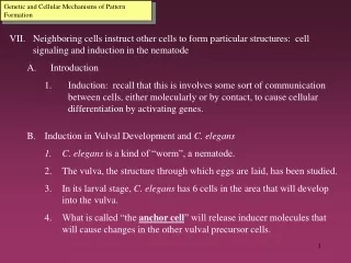
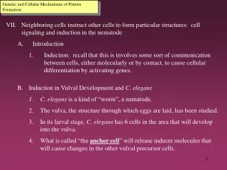
Genetic and Cellular Mechanisms of Pattern Formation. Neighboring cells instruct other cells to form particular structures: cell signaling and induction in the nematode Introduction

E N D
Genetic and Cellular Mechanisms of Pattern Formation • Neighboring cells instruct other cells to form particular structures: cell signaling and induction in the nematode • Introduction • Induction: recall that this is involves some sort of communication between cells, either molecularly or by contact, to cause cellular differentiation by activating genes. • Induction in Vulval Development and C. elegans • C. elegans is a kind of “worm”, a nematode. • The vulva, the structure through which eggs are laid, has been studied. • In its larval stage, C. elegans has 6 cells in the area that will develop into the vulva. • What is called “the anchor cell” will release inducer molecules that will cause changes in the other vulval precursor cells.
Genetic and Cellular Mechanisms of Pattern Formation • Induction in Vulval Development and C. elegans (cont’d) • Receptors in the vulva precursor cells bind to the inducer. • The cell closest to the anchor cell, receiving the highest amount of inducer, divides and differentiates to form the inner part of the vulva and also produces a second inducer. • Other vulval precursor cells have receptors for this second inducer, bind to the inducer, divide and become outer vulva cells. • This cascade of induction can be found in the formation of many other organs, in many other animals, all by signal transduction pathways.
Figure 21.17 Cell signaling and induction in the development of the nematode vulva
Genetic and Cellular Mechanisms of Pattern Formation • Programmed Cell Death (Apoptosis) • Definition: timely cell suicide during organismal development • Suicide proteins are produced that cause the cells to breakdown, die and then are engulfed by neighboring cells. • Genes and Proteins Involved in C. elegans • In C. elegans there are two apoptosis genes, ced-3 and ced-4, coding for proteins Ced-3 and Ced-4. These proteins are kept in the inactivated form in the cell (so they are stored) • Ced-9 regulates Ced-3 and Ced-4. • These proteins all activate nucleases and proteases
Genetic and Cellular Mechanisms of Pattern Formation • Programmed Cell Death (cont’d) • Apoptosis in Humans • It is thought that apoptosis proteins cause the mitochondrial membrane to leak which releases other self-destructive proteins. • One such molecule is cytochrome c which is part of the electron transport chain. • Important in: • Formation of fingers and toes • Normal operation of the immune system when cells kill viral infected cells.
Genetic and Cellular Mechanisms of Pattern Formation • Plant development depends on cell signaling and transcriptional regulation • Introduction • Cell signaling could not rely on cellular movement because of the rigid cell walls. • Plant morphogenesis or the taking of shape relies on the plane in which a cell divides. • Plants are similar to animals in relying on induction and regulating transcription. • Arabidopsis has been the main plant used for these studies.
Genetic and Cellular Mechanisms of Pattern Formation • Cell Signaling in Flower Development • Plants will develop flowers at certain times based on temperature, length of night and so these environmental cues affect cell signaling. • A floral meristem: this is a growing region of a plant that will develop into a flower. • The floral meristem has 3 layers to it. • The flower that will be produced has very different parts: petals, egg-containing carpels, stamens which bear the anthers with pollen and usually green sepals which cover the petals when the flower is closed. • Researchers found that chimeras, organisms that were made of genetically different cells, like one of the cell layers was from one plant and another cell layer from another, had floral development controlled by one of the cell layers (layer 3 or the innermost)
Genetic and Cellular Mechanisms of Pattern Formation • Organ Identity Genes in Plants (Arabidopsis) • Organ Identity Genes: these genes determine whether a petal or a stamen or some other organ will grow. • Mutations in an OIG can produce a petal where some other structure should be located. These OIG therefore are analogous to the homeotic genes in the fruit fly. • These OIG could be controlling a whole series of other genes to bring about the flower’s proper structure. • OIGs encode for transcription factors that bind to DNA
Figure 21.20a Organ identity genes and pattern formation in flower development: Normal flower development
Figure 21.20b Organ identity genes and pattern formation in flower development: In situ hybridization
Figure 21.20c Organ identity genes and pattern formation in flower development: Organ identity mutants Last Lecture Slide
Figure 21.9 Determination and differentiation of muscle cells (Layer 1)
Figure 21.9 Determination and differentiation of muscle cells (Layer 2)
Figure 21.9 Determination and differentiation of muscle cells (Layer 3)
Figure 21.15 Homologous genes that affect pattern formation in a fruit fly and a mouse