Modelling Cell Signalling and Pattern Formation
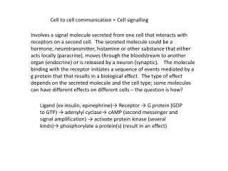
Modelling Cell Signalling and Pattern Formation. Nick Monk Department of Computer Science. Erik Plahte & Siren Veflingstad Agricultural University of Norway, Ås. Collaboration:. WTEC Systems Biology Study Group, 8 July 2004. Delta-Notch Signalling: the Neurogenic Network.

Modelling Cell Signalling and Pattern Formation
E N D
Presentation Transcript
Modelling Cell Signalling and Pattern Formation Nick Monk Department of Computer Science Erik Plahte & Siren Veflingstad Agricultural University of Norway, Ås Collaboration: WTEC Systems Biology Study Group, 8 July 2004
Delta-Notch Signalling: the Neurogenic Network Meir et al., Current Biology 12, 778-786 (2002).
Intercellular Signalling Networks • Model as a regular lattice of cells (epithelial sheet) • Intercellular signalling couples gene-protein interaction networks within each cell • May be diffusive or juxtacrine signalling
Questions and Issues • What limitations and possibilities result from patterning on cellular arrays? • At what level of detail do we have to model the internal dynamics of cells in order to understand patterning in tissues? Cells are not well-stirred bags of chemicals; internal structure is important. • How important are transient dynamics? • In development, patterning is hierarchical. Patterns are not formed from homogeneity, but from earlier patterns. Can we incorporate this in our models? • What features of intercellular signalling networks ensure that robustness and regulative capacity emerge at the tissue/organism level?
Stochastic fate assignment Loss of key genes (e.g. Dl and N) leads to over-assignment of bristles Dl and N expressed uniformly during assignment Uniform unregulated overexpression of Dl or N has little effect Delta, Notch,… Lateral Inhibition: Bristle Spacing Renaud & Simpson, Dev. Biol. 240, 361–376 (2001).
Delta-Notch–mediated cell competition Dl activates N on neighbouring cells (signalling) N activity represses Dl “activity” (within the same cell) N activity determines cell fate (via regulated transcription) D1 N2 N1 D2 Di = average of D in cells neighbouring cell i f and g are increasing and decreasing, resp. and are 1st order degradation rates
Typical Behaviour: Lateral Inhibition Collier et al., J. theor. Biol. 183, 429–446(1996).
Eukaryotic transcription and time delays • There is an irreducible delay of ~15–20 min from initiation of a transcript to appearance of functionalmRNA in the cytoplasm • The delay can be much longer (>16 hrs for human dystrophin) • Delay equations should be used to model transcription
(or distributed delay equivalents) Delayed Delta-Notch cell competition To account for the three transcriptional steps in the neurogenic network, a delay (of around an hour) should be incorporated in the competition model (Delta alone takes ~20 min to transcribe). Deal first with the simple model to assess the effect of the delay. D1 N2 N1 D2
Discrete vs. distributed delay Fixed: = 100 Distributed: = 100 +/– 32 = 3.5 Oscillations and spatial patterns
Bad news for the neurogenic network model “best case” scenario: growth of pattern from homogeneous steady state (hss). One cell on each side of hss. If non-delayed model takes 2 hours to pattern, the model with a 1 hour delay takes ca. 14 hours. More generally, the time taken to pattern grows rapidly with the delay.
Phase locking and spatial patterning Spatially inhomogeneous initial conditions can lead to blocks of phase-locked cells, separated by sharp boundaries (which can act as centres of spatial pattern formation) [c.f. somitogenesis]
Hes1 oscillates in cultured mouse cells hes1 Hes1 mRNA – Hes1 protein Half–lives: mRNA: ~24 min protein: ~22 min … but a non-delayed (ODE) model can’t oscillate… Hirata et al. predicted extra components in the feedback loop. (c.f. physics/engineering) Hirata et al., Science 298, 840–843(2002).
= 18.5 min Delay model for the Hes1 feedback loop x hes1 Hes1 mRNA y Hes1 protein The transcriptional delay has been observed directly for Hes7 Bessho et al.Genes & Dev. 17, 1451 (2003).
P53–mdm2 feedback loop x p53 z Mdm2 y mdm2 mdm2 Bar-Or et al., PNAS 97, 11250–11255(2000). [predicted “factor X” in loop using ODE model]
Hoffmann et al., Science 298, 1241–1245(2002) Nuclear NF-B – IB feedback loop Signal (e.g. TNF) NF-B IB IB nucleus IB IB note that the constitutive inhibitors (IB and IB) damp the oscillations
Conclusions • It is important to treat cells seriously in models of pattern formation. • Time delays and spatial heterogeneity can be critical. • Transcription networks involve significant delays: these affect parameter fitting, dynamics and network prediction. • The N Dl interaction is unlikely to be mediated by transcription (during competition). Post-translational protein–protein interaction? Suggest that (de novo) pattern formation is a 2–step process: 1. labile patterning by protein–protein interaction 2. fixation by regulation of gene expression.