Download
1 / 66
670 likes | 835 Vues
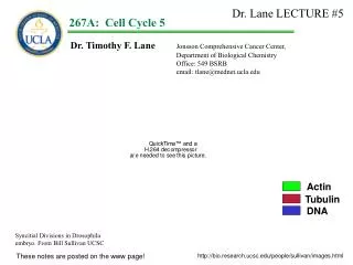
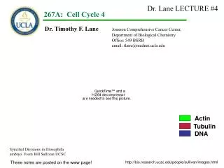
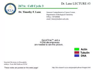
Dr. Lane LECTURE #3. 1. 267A: Cell Cycle 3. Dr. Timothy F. Lane Jonsson Comprehensive Cancer Center, Department of Biological Chemistry Office: 549 BSRB email: tlane@mednet.ucla.edu. Actin. Tubulin. DNA. Syncitial Divisions in Drosophila embryo. From Bill Sullivan UCSC.

E N D
Dr. Lane LECTURE #3 1 267A:Cell Cycle 3 Dr. Timothy F. Lane Jonsson Comprehensive Cancer Center, Department of Biological Chemistry Office: 549 BSRB email: tlane@mednet.ucla.edu Actin Tubulin DNA Syncitial Divisions in Drosophila embryo. From Bill Sullivan UCSC These notes are posted on the www page! http://bio.research.ucsc.edu/people/sullivan/images.html
Last time: We talked more about the regulation of DNA synthesis: Rao and Johnson used heterokaryons to demonstrate i. That DNA synthesis starts in response to a positive, factor that is present in S phase cells. ii. That G2 cells ignore the factor iii. That M phase cells also express a dominant chromatin condensation signal. We started our discussion of Cyclins and Cdks: i. Zellig and Langan’s work on histone kinase. Histones are sequentially phosphorylated in G1-S,G2 and then dephosphorylated in M. ii. Tim Hunts work that lead to the discovery of cyclin B in sea urchins. iii. The work of Hartwell (Sc) and Nurse (Sp) to identify cdc mutants in yeast. The cloning of yeast cdc2/cdc28 and human p34/cdc2 as the first cyclin dependent kinase. iv. The work of Draetta and Beach showing p34/p62 (cdk/cyclin) hetero dimers and how p62 stimulated p34 kinase activity. v. The work demonstrating the involvement of multiple cyclins in yeast and multiple cdks and cyclins in higher eukaryotic systems.
Goals: Identify some additional cdk targets. Examine how cdk targets regulate G1/S transitions -Regulation of Restriction Point/START -Regulation of S phase entry
G1 cyclin Yeast: cdc2 Cyclin B M G1 cdc2 G2 S cyclin cdc2 S
G1 cyclin HIGHER EUKARYOTES: cdkx Cyclin B M G1 cdc2 G2 S cyclin cdky S
RB RB Identification of cdk substrates (cont.) Regulation of RP/START and the G1/S Transition: A CDK reaction is required for human cells to traverse the G1/S boundary. The commitment to progression through the cell cycle, requires activation of E2F. At G1/S E2F family members are necessary to transcribe genes whose products are needed for DNA synthesis and other aspects of progression. In early G1, E2F is bound to another protein (the restriction boundary protein, or RB protein) and E2F is in-active. RB protein is not phosphorylated in non-dividing, G0-arrested cells, or in cells emerging into G1 from M. Cdk dependent phosphorylation of RB releases E2F from the RB/E2F complex, and permits E2F to activate transcription The CDK4/cyclin D1 complex is the major RB kinase in early-mid G1, (BUT cdk1 is sufficient1) Growth Factors R.P. Go G1 M c E2F Cyclin D1 G2 E2F CDK 4 S RP = Restriction Point/START 1Santamaría et al 2007448(7155):811-5.
The G1/S Transition As cells commit to another cell cycle, they activate members of the E2F family of transcription factors. E2F-induced genes are required for DNA synthesis. E2F Late G1
RB The G1/S Transition E2F proteins are “normally” held in an inactive state. The RB protein binds to E2F transcription factors in G0and early G1 and prevents transcription of genes for the G1 to S transition. “Restriction boundary” protein E2F Go, Early G1
RB RB The G1/S Transition Mitogen stimulation of Go/G1 cells relieves repression of transcription factors that modulate the transition into S phase. Mitogen signal for G0 cells Cell cycle progression P E2F E2F Go, Early G1 Late G1, Early S
c RB RB Cyclin D1 p The G1/S Transition Mitogen stimulation activates cdk4/cycD1 activity and phosphorylation of RB. Mitogen signal for G0 cells Cell cycle progression CDK 4 E2F E2F Go, Early G1 Late G1, Early S c-myc, cyclin E, DNApol alpha
The G1/S Transition Identification of E2F targets: Among the genes transcriptionally regulated by E2Fs: - c-myc, - cyclin E (necessary to initiate DNA synthesis), - B-myb, - p107, pRB , - E2F-1, E2F-2, - cyclin A, - p34cdk2, - DNA polymerase alpha, - dihydrofolate reductase, - thmidine kinase, - thymidylate sythase, - PCNA, RRM2 and Histone 2A. Growth Factors R.P. Go G1 M G2 S Lavia and Jansen-Durr, 1999, Bioessays 23: 221
Identification of cdk substrates: Mitogen signal for G0 cells Cyclin D1 cdk4 R.P. Go G1 Cyclin B M cdc2 G2 S
c RB RB RB Cyclin D1 p The RB protein must then be “dephosphorylated” for cells to leave M. CDK 4 E2F E2F Go, Early G1 Late G1, Early S
Identification of cdk substrates: Recall histones are transcribed only during the S phase. How is this controlled? Each mammalian histone had a distinct promoter, with different cis-acting sequences. H1 H2A H2B
Identification of cdk substrates: Recall histones are transcribed only during the S phase. How is this controlled? Each mammalian histone has a distinct promoter, with different cis-acting sequences. Importantly, Cyclin E expression is induced in response to E2F activation! Cyclin E/cdk2 dimer is required for S phase transitions. Cyclin E Cdk2 H1 H2A H2B
Identification of cdk substrates: Recall histones are transcribed only during the S phase. How is this controlled? Each mammalian histone has a distinct promoter, with different cis-acting sequences. Importantly, Cyclin E expression is induced in response to E2F activation! Cyclin E/cdk2 dimer is required for S phase transitions. Ed Harlow’s group identified NPAT (nuclear protein mapped to the AT locus) as a protein that interacts with, and is phosphorylated by, Cdk2/cyclin E1. Cyclin E Cdk2 NPAT +ATP H1 H2A H2B P NPAT P 1Zhao et al, Genes & Dev. 12, 456-461(1998)
Identification of cdk substrates: Recall histones are transcribed only during the S phase. How is this controlled? Each mammalian histone had a distinct promoter, with different cis-acting sequences. Importantly, Cyclin E expression is induced in response to E2F activation! Cyclin E/cdk2 dimer is required for S phase transitions. Ed Harlow’s group identified NPAT (nuclear protein mapped to the AT locus) as a protein that interacts with, and is phosphorylated by, Cdk2/cyclin E1. Using antibodies to NPAT, they found that NPAT localizes to histone gene complexes2. Cyclin E also co-localizes to these sites3. Localization occurs in S phase, when histone transcription occurs! Cyclin E NPAT NPAT NPAT Cdk2 NPAT +ATP H1 H2A H2B P NPAT P 1Zhao et al, Genes & Dev. 12, 456-461(1998) 2Zhao et al, Genes & Dev. 14, 2283-2297 (2000) 3Ma et al, Genes & Dev.. 12, 2298-2313 (2000)]. (This is a particularly good paper to read!).
Identification of cdk substrates: Recall histones are transcribed only during the S phase. How is this controlled? Each mammalian histone had a distinct promoter, with different cis-acting sequences. Importantly, Cyclin E expression is induced in response to E2F activation! Cyclin E/cdk2 dimer is required for S phase transitions. Ed Harlow’s group identified NPAT (nuclear protein mapped to the AT locus) as a protein that interacts with, and is phosphorylated by, Cdk2/cyclin E1. Using antibodies to NPAT, they found that NPAT localizes to histone gene complexes2. Cyclin E also co-localizes to these sites3. Localization occurs in S phase, when histone transcription occurs! Current thinking: NPAT Complex represses transcription. Cyclin E is expressed at G1/S, interacts with Cdk2, phosphorylates NPAT at the histone gene locus. Cyclin E NPAT NPAT NPAT Cdk2 NPAT +ATP H1 H2A H2B P NPAT P 1Zhao et al, Genes & Dev. 12, 456-461(1998) 2Zhao et al, Genes & Dev. 14, 2283-2297 (2000) 3Ma et al, Genes & Dev.. 12, 2298-2313 (2000)]. (This is a particularly good paper to read!).
Identification of cdk substrates: Recall histones are transcribed only during the S phase. How is this controlled? Each mammalian histone had a distinct promoter, with different cis-acting sequences. Importantly, Cyclin E expression is induced in response to E2F activation! Cyclin E/cdk2 dimer is required for S phase transitions. Ed Harlow’s group identified NPAT (nuclear protein mapped to the AT locus) as a protein that interacts with, and is phosphorylated by, Cdk2/cyclin E1. Using antibodies to NPAT, they found that NPAT localizes to histone gene complexes2. Cyclin E also co-localizes to these sites3. Localization occurs in S phase, when histone transcription occurs! Current thinking: NPAT Complex represses transcription. Cyclin E is expressed at G1/S, interacts with Cdk2, phosphorylates NPAT at the histone gene locus. Phosphorylated NPAT co-ordinates the transcription of the histone gene family, either by acting as a chromatin-rearranging machine, a co-activator, etc. Cyclin E P P P NPAT NPAT NPAT Cdk2 P P P NPAT +ATP H1 H2A H2B P NPAT P 1Zhao et al, Genes & Dev. 12, 456-461(1998) 2Zhao et al, Genes & Dev. 14, 2283-2297 (2000) 3Ma et al, Genes & Dev.. 12, 2298-2313 (2000)]. (This is a particularly good paper to read!).
Transfection with an NPAT expression vector increases histone gene transcription 10 fold. NPAT an E2F target gene3 phosphorylated at multiple Cdk2/cyclinE sites essential for cell cycle progression3 overexpression stimulates S phase entry when Phosphorylated. 1Zhao et al 2000 Genes Dev. 14:2283-97 (E Harlow) 2Ye et al 2003 Mol Cell Biol. 23:8586-600 (W Harper) 3Gao et al 2003 Mol Cell Biol. 23:2821-33 (J Zhao)
Transfection with an NPAT expression vector increases histone gene transcription 10 fold. NPAT an E2F target gene3 phosphorylated at multiple Cdk2/cyclinE sites essential for cell cycle progression3 overexpression stimulates S phase entry when Phosphorylated. luciferase H4 Fold Induction H4 H4-1 Cont b-myb dhfr 1Zhao et al 2000 Genes Dev. 14:2283-97 (E Harlow) 2Ye et al 2003 Mol Cell Biol. 23:8586-600 (W Harper) 3Gao et al 2003 Mol Cell Biol. 23:2821-33 (J Zhao)
Transfection with an NPAT expression vector increases histone gene transcription 10 fold. NPAT an E2F target gene3 phosphorylated at multiple Cdk2/cyclinE sites essential for cell cycle progression3 overexpression stimulates S phase entry when Phosphorylated. luciferase H4 Fold Induction H4 H4-1 Cont b-myb dhfr Requires N-terminal domain AND cdk2 phosphorylation sites 1Zhao et al 2000 Genes Dev. 14:2283-97 (E Harlow) Ref#2 2Ye et al 2003 Mol Cell Biol. 23:8586-600 (W Harper) 3Gao et al 2003 Mol Cell Biol. 23:2821-33 (J Zhao)
CyclinA p p p How do cdk’s choose their targets? CDK2 ATP HISTONE H1 HISTONE H1- ATP p107 protein P107- ATP E2F protein E2F-
CyclinA How do cdk’s choose their targets. The x-stal structure for cycA-cdk2 was solved in 1998 A hydrophobic patch (hp) is apparent on the surface of cyclin A, and is conserved in many cyclins. Many of the substrates of cyclin A-cdk2 have the sequence RXLFG. CDK2 hp Jeffery et al 1998 Nature 376: 313 Schulman et al 1998 PNAS 95: 10453 hpm-Hydrophobic patch mutant
CyclinA • How do cdk’s choose their targets. • The x-stal structure for cycA-cdk2 was solved in 1998 • A hydrophobic patch (hp) is apparent on the surface of cyclin A, • and is conserved in many cyclins. • Many of the substrates of cyclin A-cdk2 have the sequence RXLFG. • Schulman et al thought this patch might direct substrate recognition. • Experiment: • Mutations were made in the cyclin A hydrophobic patch (mutant hpm), • complexes were purified by immunoprecipitation, and cyclin A-cdk2 interaction • and kinase activity was examined. CDK2 ATP ? HISTONE H1 hp ATP ? p107 protein ATP ? E2F protein Schulman et al 1998 PNAS 95: 10453 hpm-Hydrophobic patch mutant
CyclinAHA cdk2 w.t. hpm Histone H1 CyclinA p107 E2F How do cdk’s choose their targets. RESULTS: The wild-type and mutated complexes could both phosphorylate H1. However, cdk2 hpm could not phosphorylate RB, and has no activity on several other [cyclin A][cdk2] substrates. CDK2 Schulman et al 1998 PNAS 95: 10453 hpm-Hydrophobic patch mutant
CyclinAHA cdk2 w.t. hpm Histone H1 CyclinA p107 E2F How do cdk’s choose their targets. RESULTS: The wild-type and mutated complexes could both phosphorylate H1. However, cdk2 hpm could not phosphorylate RB, and has no activity on several other [cyclin A][cdk2] substrates. CONCLUSIONS: These data suggest that, cdk substrates are chosen by direct interaction with the cyclin associated with a particular cdk/cyclin heterodimer. CDK2 Schulman et al 1998 PNAS 95: 10453 hpm-Hydrophobic patch mutant
Last time: We talked more about the regulation of cell cycle by cdks: i. That G1 passage required cdk4/cycD phosphorylation of pRB to release E2F and activate S phase genes including cycE. ii. That S phase entry required cycE iii. Derepression Histone genes required cycE and of pNPAT We continuted our discussion of cyclins and cdk target selection: i.
THE CYCLINS AND THE CYCLIN-DEPENDENT CATALYTIC KINASE SUBUNITS ARE ALSO SUBJECT TO CYCLIC PHOSPHORYLATION Mitogen signal for G0 cells Cyclin D1 cdk4 R.P. Cyclin E Go G1 Cyclin B M cdk2 cdk2 Cyclin A G2 cdk2 S
THE CYCLINS AND THE CYCLIN-DEPENDENT CATALYTIC KINASE SUBUNITS ARE ALSO SUBJECT TO CYCLIC PHOSPHORYLATION CyclinB (a G2/M-cyclin) accumulates in late S and in G2, and complexes with the p34cdc2 protein. Cyclin B late S and in G2 R.P. Go G1 Cyclin B M cdk2 G2 S
p THE CYCLINS AND THE CYCLIN-DEPENDENT CATALYTIC KINASE SUBUNITS ARE ALSO SUBJECT TO CYCLIC PHOSPHORYLATION CyclinB (a G2/M-cyclin) accumulates in late S and in G2, and complexes with the p34cdc2 protein. However, cdc2-cycB IP’s show no H1 kinase activity in G2. Kinase activity shoots up in mitosis when cycB protein is starting to fall! RESULTS from David Beach’s lab: 1) During early G2, p34cdc2 is phosphorylated on both tyrosine and threonine. The Tyr/Thr phosphorylation occurs in the ATP binding site of p34, and inactivates the kinase 2) When cells enter mitosis, the threonine and tyrosine of the p34 kinase in the complex is dephosphorylated, and kinase activity increases dramatically. Therefore, Tyr/Thr phosphorylation negatively regulates p34/cyclin B M-phase kinase activity. Other studies suggest a distinct threonine (thr161) site on the p34cdc2 protein must be phosphorylated to activate M-phase kinase2. Cyclin B ATP cdk2 p34 HISTONE H1 HISTONE H1- + ADP 1Draetta and Beach 1988, Cell 54: 17 2Gould et al EMBO J. 10: 3297 (1991).
p THE CYCLINS AND THE CYCLIN-DEPENDENT CATALYTIC KINASE SUBUNITS ARE ALSO SUBJECT TO CYCLIC PHOSPHORYLATION CyclinB (a G2/M-cyclin) accumulates in late S and in G2, and complexes with the p34cdc2 protein. However, cdc2-cycB IP’s show no H1 kinase activity in G2. Kinase activity shoots up in mitosis when cycB protein is starting to fall! RESULTS from David Beach’s lab Tyr/thr phosphorylation negatively regulates p34/cyclin B M-phase kinase activity. Other studies suggest a distinct threonine (thr161) site on the p34cdc2 protein must be phosphorylated to activate M-phase kinase2. Cyclin B ATP cdk2 p34 HISTONE H1 HISTONE H1- + ADP 1Draetta and Beach 1988, Cell 54: 17 2Gould et al EMBO J. 1991, 10: 3297
T14Y15 T14Y15 T14Y15 cdc2 cdc2 cdc2 T161 T161 T161 p p Cyclin B Cyclin B Cyclin B is synthesized and forms a CDK heterodimer with p34 cdc2 inactive G1 S M G2 1) During early G2, p34cdc2 is phosphorylated on both T and Y. The tyrosine/threonine phosphorylation occurs in the ATP binding site of p34 and inactivates the kinase 1Draetta and Beach 1988, Cell 54: 17 2Gould et al EMBO J. 1991, 10: 3297
T14Y15 T14Y15 T14Y15 T14Y15 T14Y15 T14Y15 cdc2 cdc2 cdc2 cdc2 cdc2 cdc2 T161 T161 T161 T161 T161 T161 p p Cyclin B Cyclin B Cyclin B Cyclin B Cyclin B is synthesized and forms a CDK heterodimer with p34 cdc2 inactive G1 G1 S Why isn’t this CDK active? M G2 1) During early G2, p34cdc2 is phosphorylated on both T and Y. The tyrosine/threonine phosphorylation occurs in the ATP binding site of p34 and inactivates the kinase 1Draetta and Beach 1988, Cell 54: 17 2Gould et al EMBO J. 1991, 10: 3297
ATP kinase…. ADP T14Y15 T14Y15 T14Y15 T14Y15 T14Y15 T14Y15 T14Y15 T14Y15 T14Y15 cdc2 cdc2 cdc2 cdc2 cdc2 cdc2 cdc2 cdc2 cdc2 T161 T161 T161 T161 T161 T161 T161 T161 T161 p p p p Cyclin B Cyclin B Cyclin B Cyclin B Cyclin B Cyclin B inactive G1 G1 G1 S Phosphorylation At T14/Y15 inhibits the activity of the CDK M G2 1) During early G2, p34cdc2 is phosphorylated on both T and Y. The tyrosine/threonine phosphorylation occurs in the ATP binding site of p34 and inactivates the kinase 1Draetta and Beach 1988, Cell 54: 17 2Gould et al EMBO J. 1991, 10: 3297
ATP kinase…. ADP T14Y15 T14Y15 T14Y15 T14Y15 T14Y15 T14Y15 T14Y15 T14Y15 T14Y15 T14Y15 T14Y15 T14Y15 T14Y15 cdc2 cdc2 cdc2 cdc2 cdc2 cdc2 cdc2 cdc2 cdc2 cdc2 cdc2 cdc2 cdc2 T161 T161 T161 T161 T161 T161 T161 T161 T161 T161 T161 T161 T161 p p p p p p p Cyclin B Cyclin B Cyclin B Cyclin B Cyclin B Cyclin B Cyclin B Cyclin B Cyclin B inactive G1 G1 G1 G1 S (inactive) M G2 (still inactive) 1) During early G2, p34cdc2 is phosphorylated on both T and Y. The tyrosine/threonine phosphorylation occurs in the ATP binding site of p34 and inactivates the kinase 1Draetta and Beach 1988, Cell 54: 17 2Gould et al EMBO J. 1991, 10: 3297
ATP kinase…. ADP T14Y15 T14Y15 T14Y15 T14Y15 T14Y15 T14Y15 T14Y15 T14Y15 T14Y15 T14Y15 T14Y15 T14Y15 T14Y15 T14Y15 T14Y15 T14Y15 T14Y15 T14Y15 T14Y15 T14Y15 T14Y15 T14Y15 T14Y15 cdc2 cdc2 cdc2 cdc2 cdc2 cdc2 cdc2 cdc2 cdc2 cdc2 cdc2 cdc2 cdc2 cdc2 cdc2 cdc2 cdc2 cdc2 cdc2 cdc2 cdc2 cdc2 cdc2 T161 T161 T161 T161 T161 T161 T161 T161 T161 T161 T161 T161 T161 T161 T161 T161 T161 T161 T161 T161 T161 T161 T161 p p p p p p p p Cyclin B Cyclin B Cyclin B Cyclin B Cyclin B Cyclin B Cyclin B Cyclin B Cyclin B Cyclin B Cyclin B Cyclin B Cyclin B Cyclin B Cyclin B Cyclin B Cyclin B PPase inactive G1 G1 G1 G1 G1 G1 S S S (inactive) M M M G2 G2 G2 (still inactive) ATP Active cdc2!! T14 dephosphorylation Y15 dephosphorylation When cells enter mitosis, the T and Y of the p34 kinase in the complex are dephosphorylated, and kinase activity increases dramatically. 1Draetta and Beach 1988, Cell 54: 17 2Gould et al EMBO J. 1991, 10: 3297
ATP kinase…. ADP T14Y15 T14Y15 T14Y15 T14Y15 T14Y15 T14Y15 T14Y15 T14Y15 T14Y15 T14Y15 T14Y15 T14Y15 T14Y15 T14Y15 T14Y15 T14Y15 T14Y15 T14Y15 T14Y15 T14Y15 T14Y15 T14Y15 T14Y15 cdc2 cdc2 cdc2 cdc2 cdc2 cdc2 cdc2 cdc2 cdc2 cdc2 cdc2 cdc2 cdc2 cdc2 cdc2 cdc2 cdc2 cdc2 cdc2 cdc2 cdc2 cdc2 cdc2 T161 T161 T161 T161 T161 T161 T161 T161 T161 T161 T161 T161 T161 T161 T161 T161 T161 T161 T161 T161 T161 T161 T161 p p p p p p p p Cyclin B Cyclin B Cyclin B Cyclin B Cyclin B Cyclin B Cyclin B Cyclin B Cyclin B Cyclin B Cyclin B Cyclin B Cyclin B Cyclin B Cyclin B Cyclin B Cyclin B PPase inactive G1 G1 G1 G1 G1 G1 S S S (inactive) M M M G2 G2 G2 (still inactive) ATP Active cdc2!! T14 dephosphorylation Y15 dephosphorylation Other studies suggest a distinct (thr161) site on the p34cdc2 protein must be P to activate M-phase kinase2 1Draetta and Beach 1988, Cell 54: 17 2Gould et al EMBO J. 1991, 10: 3297
ATP kinase…. ADP T14Y15 T14Y15 T14Y15 T14Y15 T14Y15 T14Y15 T14Y15 T14Y15 T14Y15 T14Y15 T14Y15 T14Y15 T14Y15 T14Y15 T14Y15 T14Y15 T14Y15 T14Y15 T14Y15 T14Y15 T14Y15 T14Y15 T14Y15 cdc2 cdc2 cdc2 cdc2 cdc2 cdc2 cdc2 cdc2 cdc2 cdc2 cdc2 cdc2 cdc2 cdc2 cdc2 cdc2 cdc2 cdc2 cdc2 cdc2 cdc2 cdc2 cdc2 T161 T161 T161 T161 T161 T161 T161 T161 T161 T161 T161 T161 T161 T161 T161 T161 T161 T161 T161 T161 T161 T161 T161 p p p p p p p p Cyclin B Cyclin B Cyclin B Cyclin B Cyclin B Cyclin B Cyclin B Cyclin B Cyclin B Cyclin B Cyclin B Cyclin B Cyclin B Cyclin B Cyclin B Cyclin B Cyclin B PPase inactive G1 G1 G1 G1 G1 G1 Cycin B is degraded S S S (inactive) M M M G2 G2 G2 (still inactive) ATP Active cdc2!! T14 dephosphorylation Y15 dephosphorylation
THE WHAT ENZYMES ARE RESPONSIBLE FOR THE P AND de P OF THE p34cdc2 IN THE CELL CYCLE? Yeast studies have identified several regulators of p34/cyclin complex activity. wee1, mik1, and cdc25: regulate Cdc2 kinase activity at G2/M wee1 cdc 25 Cdc2 p34 Mik 1 cdc13 (cyclin) Interphase mitosis G2 M In the absence of both wee1 and mik1 cells can enter mitosis prematurely, without completing DNA synthesis, because they have too much CDK activity!
p p THE WHAT ENZYMES ARE RESPONSIBLE FOR THE P AND de P OF THE p34cdc2 IN THE CELL CYCLE? Yeast studies have identified several regulators of p34/cyclin complex activity. wee1, mik1, and cdc25: regulate Cdc2 kinase activity at G2/M wee1 is a both a Y and a T/S kinase cdc2-cdc13 heterodimers are the substrate Phosphorylation of p34cdc2/cyclin B BLOCKS H1 kinase activity! wee1 ADP Cdc2 p34 cdc13 (cyclin B) ATP HISTONE H1 pH1 1Parker et al, 1992 PNAS 89: 2917
T14Y15 T14Y15 T14Y15 T14Y15 T14Y15 T14Y15 T14Y15 T14Y15 T14Y15 T14Y15 T14Y15 T14Y15 T14Y15 T14Y15 T14Y15 T14Y15 T14Y15 T14Y15 T14Y15 T14Y15 T14Y15 T14Y15 T14Y15 cdc2 cdc2 cdc2 cdc2 cdc2 cdc2 cdc2 cdc2 cdc2 cdc2 cdc2 cdc2 cdc2 cdc2 cdc2 cdc2 cdc2 cdc2 cdc2 cdc2 cdc2 cdc2 cdc2 T161 T161 T161 T161 T161 T161 T161 T161 T161 T161 T161 T161 T161 T161 T161 T161 T161 T161 T161 T161 T161 T161 T161 p p p p p p p p Cyclin B Cyclin B Cyclin B Cyclin B Cyclin B Cyclin B Cyclin B Cyclin B Cyclin B Cyclin B Cyclin B Cyclin B Cyclin B Cyclin B Cyclin B Cyclin B Cyclin B PPase ATP THE WHAT ENZYMES ARE RESPONSIBLE? Wee1 ADP inactive G1 G1 G1 G1 G1 G1 Cycin B is degraded S S S (inactive) M M M G2 G2 G2 (still inactive) ATP Active cdc2!! T14 dephosphorylation Y15 dephosphorylation
wee1 gene: The human wee1 gene was cloned by rescuing a yeast wee1 mutant with a human cDNA expression library1. wee1 cooperates with Mik1, also a protein tyrosine kinase2! wee1 and mik1 are essential for p34cdc2 phosphorylation. wee1 cdc 25 Cdc2 p34 Mik 1 cdc13 (cyclin) Overexpression of human wee1 in HeLa cells prevents cell cycle progression3. Suggest that Wee1 must be inactivated for a cell to make a transition into M4. Interphase mitosis G2 M Watanabe et al demonstrated hyperphosphorylation and subsequent degradation of Wee1 in HeLa cells4. 1Igarashi et al, 1991 Nature 353: 80 2Lundgren et al, 1991, Cell 64:1111 3McGowan and Russell 1993, EMBO J 12:75. 4Watanabe et al 1995, EMBO J 14:1878
p p p p PROTEIN PHOSPHATASES THAT REGULATE THE M-PHASE KINASE: Histone Histone P34 cdc2 cdc13 ?? Wee 1 P34 cdc2 cdc13 Histone Histone Strausfeld et al, 1991, Nature 351: 242 Gautier et al, 1991, Cell 67; 197.
p p p p PROTEIN PHOSPHATASES THAT REGULATE THE M-PHASE KINASE: The yeast cdc25 gene product is a protein Y and T phosphatase - highly specific for tyrosine-phosphorylated p34cdc2. - can also dephosphorylate the adjacent threonine residue; Dephosphorylation of p34cdc2 protein activates the p34/cyclin kinase activity Histone Histone P34 cdc2 cdc13 Wee 1 Cdc 25 P34 cdc2 cdc13 Histone Histone Strausfeld et al, 1991, Nature 351: 242 Gautier et al, 1991, Cell 67; 197.
PROTEIN PHOSPHATASES THAT REGULATE THE M-PHASE KINASE: The cdc25 gene product, a cdc2 P’tase, appears to be expressed primarily in G2 both in S. pombe1 and in HeLa cells2 Hela Cells, analysis by elutriation centrifugation cdc25 Several mammalian homologues of Cdc25 have now been cloned. Cdc25C can dephosphorylate p34cdc2, the G2 Cdk. 1Moreno et al 1990, Nature 344: 549 2Sadhu et al 1990 PNAS 87: 5139
Cdc 25 Cdc 25 cdc2 cdc2 cdc2 Cyclin Cyclin Cyclin B B B p p p p p PROTEIN PHOSPHATASES THAT REGULATE THE M-PHASE KINASE: Phosphorylation activates the phosphatase activity of the Cdc25 protein. The Cdc2 protein component of the Cdc2/cyclin B complex (the “histone kinase”) must be dephosphorylated at residues 14 and 15. Wee 1 Histone + ATP This sets up a cycle in which rapid activation of p34cdc2/ cyclin B histone kinase. Histone Hoffmann et al, 1993, EMBO J. 12:53.
Cdc 25 Cdc 25 cdc2 cdc2 cdc2 Cyclin Cyclin Cyclin B B B p p p p p PROTEIN PHOSPHATASES THAT REGULATE THE M-PHASE KINASE: Phosphorylation activates the phosphatase activity of the Cdc25 protein. The Cdc2 protein component of the Cdc2/cyclin B complex (the “histone kinase”) must be dephosphorylated at residues 14 and 15. Cdc2/cyclin B histone kinase can also phosphorylate the Cdc25 phosphatase Phosphorylation of Cdc25 activates this cdc25. Thus a "self-amplification" is set up. PPase Wee 1 Histone + ATP MO15 CycH Histone
T14Y15 T14Y15 T14Y15 T14Y15 T14Y15 T14Y15 T14Y15 T14Y15 T14Y15 T14Y15 T14Y15 T14Y15 T14Y15 T14Y15 T14Y15 T14Y15 T14Y15 T14Y15 T14Y15 T14Y15 T14Y15 T14Y15 T14Y15 cdc2 cdc2 cdc2 cdc2 cdc2 cdc2 cdc2 cdc2 cdc2 cdc2 cdc2 cdc2 cdc2 cdc2 cdc2 cdc2 cdc2 cdc2 cdc2 cdc2 cdc2 cdc2 cdc2 T161 T161 T161 T161 T161 T161 T161 T161 T161 T161 T161 T161 T161 T161 T161 T161 T161 T161 T161 T161 T161 T161 T161 p p p p p p p p Cyclin B Cyclin B Cyclin B Cyclin B Cyclin B Cyclin B Cyclin B Cyclin B Cyclin B Cyclin B Cyclin B Cyclin B Cyclin B Cyclin B Cyclin B Cyclin B Cyclin B PPase ATP MO15 CycH Wee1 ADP inactive G1 G1 G1 G1 G1 G1 Cyclin B is degraded S S S A cdk also regulates cdk2 creating a cdk cascade. p34 cdc2/28 must be phosphorylated on Thr161 by cdc2-activating Kinase, or CAK. CAK is composed of a cdk called MO15 and a new G1 cyclin, called cyclin H. CAK can activate both Cdc2 (a G2/M Cdk) and Cdk2 (a G1 Cdk). (inactive) M M M G2 G2 G2 (still inactive) ATP Active CDK!! T14 dephosphorylation Y15 dephosphorylation Fisher and Morgan, 1994, Cell 78: 713
NEGATIVE REGULATION OF THE CELL CYCLE, CYCLINS AND CDKS: How do external agents interfere with the sequential cyclin/cdk activations, and regulate progression through the cycle?