Download
1 / 65
670 likes | 1.01k Vues
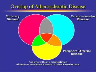
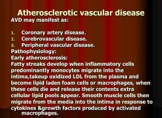
Atherosclerotic vascular disease. AVD may manifest as: Coronary artery disease. Cerebrovascular disease. Peripheral vascular disease. Pathophysiology : Early atherosclerosis: Fatty streaks develop when inflammatory cells predominantly monocytes migrate into the

E N D
Atherosclerotic vascular disease AVD may manifest as: Coronary artery disease. Cerebrovascular disease. Peripheral vascular disease. Pathophysiology: Early atherosclerosis: Fatty streaks develop when inflammatory cells predominantly monocytes migrate into the intima,takeup oxidized LDL from the plasma and become lipid laden foam cells or macrophages, when these cells die and release their contents extra cellular lipid pools appear. Smooth muscle cells then migrate from the media into the intima in response to cytokines &growth factors produced by activated macrophages.
The lipid core will be covered by smooth muscle cells and matrix, producing a stable atherosclerotic plaque which is asymptomatic until it becomes large enough to obstruct arterial flow. Advanced atherosclerosis: • In an established atherosclerotic plaque macrophages mediate inflammation & smooth muscle cells promote repair, if inflammation predominates, the plaque becomes active or unstable & may be complicated by ulceration & superadded thrombosis. Cytokines such as interleukin-1, tumour necrosis-alpha, interferon gamma, platelet derived growth factors, and matrix metalloprotinases are released by activated macrophages & may cause the initial smooth muscle
Cells overlying the plaque to become senescent resulting in thinning of the protective fibrous cap, may lead to erosion, fissuring or rupture of the plaque surface, exposing its content to circulating blood, & may trigger platelet aggregation & thrombosis, may cause partial or complete obstruction resulting in infarction or ischemia of the affected organ.
Risk Factors A. Fixed R. F • Age • Male sex • Family history. B. Modifiable R.F • Smoking - Sedentary lifestyle • Hypertension - Obesity • Lipid disoder. - Diet • D.M • Haemostatic variables
Primary Prevention Population advice to prevent coronary disease; • Do not smoke. • Take regular exercise (minimum of 20 minutes three times a week). • Maintain ideal body weight . • Eat a mixed diet rich in fresh fruit & vegetables. • Aim to get no more than 30% of energy intake from saturated fat.
Secondary prevention: • Patients who already have evidence of atheromatous vascular disease e.g M.I, are at high risk of another vascular event, can be offered a variety of treatment and measures to improve their outlook (secondary prevention). • Correction of risk factors • Aspirin • Beta blockers • ACE Inhibitors
Coronary Heart Disease • It is the most common form of heart disease. • Most important cause of premature death. • In UK 1 in 3 men & 1 in 4 women die of CHD. Clinical manifestation: • Stable angina. • Unstable angina. • M.I • Heart failure. • Arrhythmia. • Sudden death.
Stable angina • Angina pectoris is caused by transient myocardial ischemia. • It may occur whenever there is an imbalance between myocardial oxygen supply & demand. • Coronary atheroma is the most common cause. • Other causes include aortic valve disease & hypertrophic cardiomyopathy.
Clinical features: • The history is the most important factor in making the diagnosis. • Central chest pain, discomfort or breathlessness that is precipitated by exertion or other forms of stress, (heavy meal, cold exposure, intense emotion, lying flat/ decubitus, vivid dreams/ nocturnal angina), and relieved by rest. • Start up angina: the pain comes when they start walking & that later it dose not return despite greater effort. • Physical examination is frequently negative, but should include: • Evidence of valve disease, important risk factors, left ventricular dysfunction, features of arterial disease e.g carotid bruits and untreated conditions that may exacerbate angina e.g anemia, thyrotoxicosis.
Investigations: Resting ECG: • Evidence of previous M.I, but may be normal. • Occasionally T wave flattening or inversion. • The most important ECG changes is reversible ST segment depression or elevation with or without T wave inversion at the time the patient is experiencing symptoms. Exercise ECG: • Standard treadmill or bicycle ergometer protocol, planar or down-sloping ST-segment depression of 1 mm or more is indicative of ischemia, up-sloping is less specific & often occurs in normal individuals. • Exercise ECG is used to diagnose angina, assess severity & identifying high- risk individuals. • False +ve: Digoxin, LVH, LBBB, WPW syndrome, the accuracy is lower in women than men.
Risk stratification in stable angina. • High Risk; *post –infarct angina, *poor effort tolerance, *ischemia at low workload, *left main or three vessel disease, *poor LV function. Low Risk: • Predictable exertional angina, *good effort tolerance, *ischemia only at high workload, *single-vessel or minor two-vessel disease, *good LV function. Other forms of stress testing: • Myocardial perfusion scanning: useful when exercise test is not diagnostic, or patient can not exercise, its accuracy is higher than exercise test. The technique involve obtaining scintiscans of the myocardium at rest and during stress after administration of an i.v radioactive isotope e.g thallium201, it is taken up by viable myocardium.
A perfusion defect present during stress but not rest indicates reversible myocardial ischemia, whereas persistent perfusion defect indicates previous M.I Stress echocardiography: It is alternative to perfusion scanning with similar accuracy. Uses transthoracic echo to identify ischemic segments of myocardium & area of infarction, the latter do not contract at rest or during stress. Coronary arteriography: Shows the extent & nature of coronary artery disease, it may be indicated when other investigations fail to diagnose the cause of atypical chest pain.
Management • Assessment of the extent & severity of arterial disease. • Identification & control of significant risk factors • Measures to control symptoms. • Identification of high risk patients & application of treatment to improve life expectancy. Antiplatelet therapy: Aspirin 75-150 mg reduce the risk of MI, Clopidogrel 75 mg daily equally effective but more expensive can be used if the patient has dyspepsia.
Anti-anginal drugs: • Nitrates: produces venous & arteriolar dilatation • Decrease myocardial oxygen demand (lower preload & afterload) & increase myocardial oxygen supply. • Sublingual glyceryl trinitrate (GTN), as aerosol 400 microgm or tablet 300-500 micro-gm sublingually usually relieve angina in 2-3 minutes, side- effects include headache, symptomatic hypotension & syncope, the tablet should be replaced 8 weeks after the bottle has been opened. • Nitrates can be used prophylactically before exercise. • GTN is subject to extensive first pass metabolism in the liver, its ineffeective when swallowed. • Nitrate free period of 6-8 hr. every day to avoid tolerance.
Preparation Peak actionDuration Sublingual GTN 4-8 mins 10-30 mins Buccal GTN 4-10 mins 30-300 mins Transdermal GTN 1-3 hrs up to 24 hrs Oral isosorbide dinitrate 45-120 mins 2-6 hrs Oral isosorbide mononitrate 45-120 mins 6-10 hrs
Beta-blockers • Reduce myocardial oxygen demand by reducing heart rate, BP, and myocardial contractility. • Non-selective BB may exacerbate coronary spasm by blocking Beta 2 coronary adrenoceptors. • Give once daily cardioselective preparation, atenolol 50-100 mg daily, slow release metoprolol 200 mg daily, bisoprolol 5-10 mg daily. • Ββ should not be withdrawn suddenly as this may cause arrhythmia, more angina or MI.(ββ withdrawal syndrome). Calcium antagonists: • lower myocardial oxygen demand by reducing blood pressure and myocardial contractility. • Dihydropyridine calcium antagonists, such as nifedipine and nicardipine, often cause a reflex tachycardia; it is often best to use these drugs in combination with a β-blocker. • In contrast, verapamil and diltiazem are particularly suitable for patients who are not receiving a β-blocker because they inhibit conduction through the AV node and tend to cause a bradycardia or even atrioventricular block in susceptible individuals.
The calcium antagonists may reduce myocardial contractility and can aggravate or precipitate heart failure. Other unwanted effects include peripheral oedema, flushing, headache and dizziness. Potassium channel activators; This class of drug has arterial and venous dilating properties but does not exhibit the tolerance seen with nitrates. Nicorandil (10-30 mg 12-hourly orally) is the only drug in this class currently available for clinical use. • it is conventional to start therapy with low-dose aspirin, sublingual GTN and a β-blocker, and then add a calcium channel antagonist or a long-acting nitrate later, if necessary.
The goal is the control of angina with minimum side-effects and the simplest possible drug regimen. There is little or no evidence that prescribing multiple anti-anginal drugs is of benefit, and revascularisation should be considered if an appropriate combination of two drugs fails to achieve a symptomatic response. • Invasive treatment The most widely used invasive options for the treatment of ischaemic heart disease include percutaneous coronary intervention (PCI; including percutaneous transluminal coronary angioplasty, PTCA) and coronary artery bypass graft (CABG) surgery.
UNSTABLE ANGINA • is a clinical syndrome that is characterised by new-onset or rapidly worsening angina (crescendo angina), angina on minimal exertion or angina at rest. • The condition shares common pathophysiological mechanisms with acute myocardial infarction, • the term 'acute coronary syndrome' is used to describe these disorders collectively. • These entities comprise a spectrum of disease that encompasses ischaemia with no myocardial damage, ischaemia with minimal myocardial damage, partial thickness (non-Q wave) myocardial infarction, and full thickness (Q wave) myocardial infarction .
An acute coronary syndrome may present as a new phenomenon or against a background of chronic stable angina. • The culprit lesion is usually a complex ulcerated or fissured atheromatous plaque with adherent platelet-rich thrombus and local coronary artery spasm . Diagnosis and risk stratification • The assessment of acute chest pain depends heavily on an analysis of the character of the pain and its associated features, evaluation of the ECG, and serial measurements of biochemical markers of cardiac damage, such as troponin I and T.
A 12-lead ECG is mandatory and is the most useful method of initial triage. • Evolving transmural infarction is characterised by persistent ST elevation, new Q waves or new left bundle branch block • In patients with unstable angina or partial thickness (non-Q wave or non-ST elevation) myocardial infarction, the ECG may show ST/T wave changes including ST depression, transient ST elevation and T-wave inversion; the T-wave changes are sometimes prolonged. • Approximately 12% of patients with well-characterised unstable angina or non-ST segment elevation myocardial infarction progress to acute infarction or death, and almost one-third will suffer a recurrence of severe ischaemic pain, within 6 months of the index event.
The risk markers that are indicative of an adverse prognosis include : • recurrent ischaemia, • extensive ECG changes at rest or during pain, • the release of biochemical markers (creatinekinase or troponin), • arrhythmias and haemodynamic complications (e.g. hypotension, mitral regurgitation) during episodes of ischaemia • those who experience unstable angina following acute myocardial infarction are also at increased risk.
Risk stratification is important because it guides the use of more complex pharmacological and interventional treatment .
High risk: Clinical: • Post-infarct anginaRecurrent pain at restHeart failure ECG: • ArrhythmiaST depressionTransient ST elevationPersistent deep T-wave inversion • Biochemistry:Troponin T > 0.1 μg/l
Low risk: Clinical • No history of MI • Rapid resolution of symptoms ECG: Minor or no ECG changes Biochemistry: • Troponin T < 0.1 microgm/ l
The initial treatment should include • bed rest, • antiplatelet therapy (aspirin 300 mg followed by 75-325 mg daily long-term and clopidogrel 300 mg followed by 75 mg daily for 12 months, • anticoagulant therapy (e.g. unfractionated or fractionated heparin) • β-blocker (e.g. atenolol 50-100 mg daily or metoprolol 50-100 mg 12-hourly • A dihydropyridine calcium antagonist (e.g. nifedipine or amlodipine) can be added to the β-blocker, but may cause an unwanted tachycardia if used alone; verapamil or diltiazem is therefore the calcium antagonist of choice if a β-blocker is contraindicated
An intravenous infusion of unfractionated heparin (with dose adjusted according to the activated partial thromboplastin time) or weight-adjusted subcutaneous low molecular weight heparin (e.g. enoxaparin 1 mg/kg 12-hourly) should be given • If pain persists or recurs, infusions of intravenous nitrates (e.g. GTN 0.6-1.2 mg/hr or isosorbide dinitrate 1-2 mg/hr) or buccal nitrates may help, but such patients should also be considered for early revascularisation. • Refractory cases or those with haemodynamic compromise should be considered for a glycoprotein IIb/IIIa receptor antagonist (e.g. abciximab, tirofiban or eptifibatide), intra-aortic balloon pump or emergency coronary angiography .
Most low-risk patients stabilise with aspirin, clopidogrel, heparin and anti-anginal therapy, and can be gradually mobilised. If there are no contraindications, • exercise testing may be performed prior to or shortly following discharge. • Coronary angiography should be considered with a view to revascularisation in all patients at moderate or high risk, including those who fail to settle on medical therapy, those with extensive ECG changes, those with an elevated plasma troponin and those with severe pre-existing stable angina. • This often reveals disease that is amenable to PCI ; however, if the lesions are not suitable for PCI the patient should be considered for urgent CABG.
MYOCARDIAL INFARCTION • Myocardial infarction (MI) is almost always due to the formation of occlusive thrombus at the site of rupture or erosion of an atheromatous plaque in a coronary artery. • The thrombus often undergoes spontaneous lysis over the course of the next few days, although by this time irreversible myocardial damage has occurred. • Without treatment the infarct-related artery remains permanently occluded in 30% of patients. • The process of infarction progresses over several hours and therefore most patients present when it is still possible to salvage myocardium and improve outcome.
CLINICAL FEATURES • Pain is the cardinal symptom of MI, but breathlessness, vomiting, and collapse or syncope are common features. • The pain occurs in the same sites as angina but is usually more severe and lasts longer; • It is often described as a tightness, heaviness or constriction in the chest. At its worst, the pain is one of the most severe which can be experienced and the patient's expression and pallor may vividly convey the seriousness of the situation • Most patients are breathless and in some this is the only symptom. Indeed, some myocardial infarcts pass unrecognised.
Painless or 'silent' myocardial infarction is particularly common in older or diabetic patients. • If syncope occurs, it is usually due to an arrhythmia or profound hypotension. Vomiting andsinus bradycardia are often due to vagal stimulation and are particularly common in patients with inferior MI. • Nausea and vomiting may also be caused or aggravated by opiates given for pain relief. • Sometimes infarction occurs in the absence of physical signs. • Sudden death, from ventricular fibrillation or asystole, may occur immediately, and many deaths occur within the first hour.
If the patient survives this most critical stage, the liability to dangerous arrhythmias remains, but diminishes as each hour goes by. • The development of cardiac failure reflects the extent of myocardial damage and is the major cause of death in those who survive the first few hours of infarction.
Physical signs • Signs of sympathetic activation • Pallor, sweating, tachycardia • Signs of vagal activation • Vomiting, bradycardia • Signs of impaired myocardial function • Hypotension, oliguria, cold peripheries • Narrow pulse pressure • Raised jugular venous pressure • Third heart sound • Quiet first heart sound • Diffuse apical impulse • Lung crepitations • Signs of tissue damage • Fever • Signs of complications, e.g. mitral regurgitation, pericarditis
INVESTIGATIONS : • Electrocardiography The ECG is usually helpful in confirming the diagnosis; however, it may be difficult to interpret if there is bundle branch block or evidence of previous MI. • Only rarely is the initial ECG entirely normal, but in up to one-third of cases the initial ECG changes may not be diagnostic. • The earliest ECG change is usually ST elevation; later on there is diminution in the size of the R wave, and in transmural (full thickness) infarction a Q wave begins to develop. • Subsequently, the T wave becomes inverted, this change persists after the ST segment has returned to normal.
In contrast to transmural lesions, partial thickness or subendocardial infarction causes ST/T wave changes, without Q waves or prominent ST elevation; this is often accompanied by some loss of the R waves in the leads facing the infarct and is also known as non-Q wave or non-ST elevation myocardial infarction. • The ECG changes are best seen in the leads that 'face' the infarcted area. • When there has been anteroseptal infarction, abnormalities are found in one or more leads from V1 to V4 • while anterolateral infarction produces changes from V4 to V6, in aVL and in lead I.
Inferior infarction is best shown in leads II, III and aVF, while at the same time leads I, aVL and the anterior chest leads may show 'reciprocal' changes of ST depression • Infarction of the posterior wall of the left ventricle does not cause ST elevation or Q waves in the standard leads, but can be diagnosed by the presence of reciprocal changes (ST depression and a tall R wave in leads V1-V4). • Some infarctions (especially inferior) also involve the right ventricle; this may be identified by recording from additional leads placed over the right precordium.
The serial evolution of ECG changes in full thickness myocardial infarction. A. Normal ECG complex. B. Acute ST elevation ('the current of injury'). C. Progressive loss of the R wave, developing Q wave, resolution of the ST elevation and terminal T wave inversion. D. Deep Q wave and T wave inversion. E. Old infarction
Plasma biochemical markers • The biochemical markers that are most widely used in the detection of MI are creatine kinase (CK), a more sensitive and cardiospecific isoform of this enzyme (CK-MB), and the cardiospecific proteins, troponins T and I • The troponins are also released, to a minor degree, in unstable angina with minimal myocardial damage Serial (usually daily) estimations are particularly helpful because it is the change in plasma concentrations of these markers that is of diagnostic value.
CK starts to rise at 4-6 hours, peaks at about 12 hours and falls to normal within 48-72 hours. • CK is also present in skeletal muscle, and a modest rise in CK (but not CK-MB) may sometimes be due to an intramuscular injection, vigorous physical exercise or, in old people particularly, a fall. • Defibrillation causes significant release of CK but not CK-MB or troponins. The most sensitive markers of myocardial cell damage are the cardiac troponins T and I, which are released within 4-6 hours and remain elevated for up to 2 weeks. • Other blood tests A leucocytosis is usual, reaching a peak on the first day. The erythrocyte sedimentation rate (ESR) becomes raised and may remain so for several days. C-reactive protein (CRP) is also elevated in acute MI.
Chest X-ray • This may demonstrate pulmonary oedema that is not evident on clinical examination The heart size is often normal but there may becardiomegaly due to pre-existing myocardial damage. • Echocardiography • This can be performed at the bedside and is a very useful technique for assessing left and right ventricular function and for detecting important complications such as mural thrombus, cardiac rupture, ventricular septal defect, mitral regurgitation and pericardial effusion.