Download
1 / 29
290 likes | 750 Vues
surgical management of UPJ-O by Dr.adah ceifo

E N D
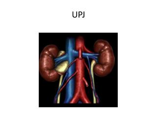
Surgical Management of UPJ-O Dr. Wadah Ceifo FA.Uro,Berlin.Germany
PATHOPHYSIOLOGY • (UPJ-o) may be defined as a functional or anatomic obstruction to urine flow from the renal pelvis to the proximal ureter that results in symptoms or renal damage. • multiple etiologic factors. • Primary, Congenital : aperistaltic segment of the ureter (Intrinsic) • extrinsic compression : kinks, bands, polar vessels, and a high insertion of the ureter may be obvious. .
Clinical Presentation and Diagnosis • UPJ obstruction is one of the most common causes of prenatal hydronephrosis. • Congenital UPJ obstruction can present at any time, from intrauterine life to old age. • In adults : flank pain ,nause, hematuria, urinary tract infections, stone disease, or vague gastrointestinal complaints. • The diagnosis of UPJ obstruction is based on the combination of clinical manifestations, radiographic evidence of obstruction and impairment of renal function • radiographic study :a diuretic intravenous pyelogram, radionuclide renal scans, computed tomography, ultrasonography, and retrograde pyelography.
SURGICALMANAGEMENT • The primary indications for treatment of UPJ obstruction include relief of pain and relief of physiologically significant obstruction. In addition, recurrent stone formation or infection may indicate the need for surgical reconstruction of the UPJ. The ultimate goal is to provide a drainage system with unobstructed urinary flow . • surgical approaches to correction of UPJ obstruction, they can be classified into 3 categories: • 1)- Open surgical procedures (pyeloplasty) • 2)- Endoscopic (antegrade or retrograde) procedures • 3)- Laparoscopic procedures. While considering these various options. • it is important to weigh the potential risks and benefits of these approaches, the success rates, and to keep in mind that long-term results are pending in some cases.
Dismembered Pyeloplasty • popularized and modified by Anderson & Hynes . • can be applied or modified to reconstruct the vast majority of UPJ obstructions. • most popular of all open procedures. • allows the excision of the anatomically strictured area. • its utilization is not dependent on whether the ureteral insertion is high or normal. • It does not provide a good result ,when there is a lengthy proximal ureteral stricture associated with a poorly accessible intrarenal pelvis.
Culp-DeWeerd Spiral Flap • The primary role of this procedure is when there is a proximal ureteral stricture associated with a UPJ obstruction. • It should be performed in the presence of a large extrarenal pelvis, as the size of the flap is limited only by the renal pelvis. • UPJ obstruction associated with high insertion of the ureter can be difficult to repair with this technique.
Foley Y-V Plasty • It was originally developed to reconstruct the obstructed system associated with a high ureteral insertion into the renal pelvis. • It is not well suited when a proximal ureteral stricture is present, where lower pole vessel transposition is indicated, or when the reduction of procedure.
Scardino-Prince Vertical Flap • It is largely of historic interest only . • Its application was limited to obstruction of an already dependent UPJ that was situated on the medial aspect of an extrarenal pelvis . • It can be used to manage proximal ureteral strictures, but it cannot provide the length and versatility of the spiral flap..
Ureterocalycostomy • Itis an important procedure in certain clinical situations. • It is most commonly employed as a salvage procedure after a failed pyeloplasty, particularly in situations where a repeat pyeloplasty will likely fail secondary to fibrosis of the renal pelvis. • It may be used as the primary reconstructive procedure for UPJ obstructions associated with rotational or fusion anomalies, such as a horseshoe kidney( allows a dependent drainage of the unit without the need to sacrifice the isthmus). • it can be utilized when a small intrarenal pelvis is present. • The critical portion is to resect sufficient lower pole parenchyma to prevent subsequent renal cortical fibrosis. .
AntegradeEndopyelotomy • a vessel (artery or vein) crossing within 1.5 cm of the PUJ; most (91%) are located anterior to the PUJ and the rest are posterior. Rarely are these vessels located lateral to the PUJ; and hence the PUJ is cut laterally • The main advantages : a small incision which results in minimal morbidity, good postoperative drainage of the kidney , surgeon is able to visualize the obstruction to avoid incising a crossing vessel. • presence of a crossing vessel lowered the success rate from 82% to 33%. • disadvantages : the need for a nephrostomy tube, the risk of bleeding with the possibility of emergency embolization. • However, the overall decreased morbidity coupled with a high success rate that has been duplicated in many large series at many institutions around the world make this procedure an excellent choice in most adult patients.
RetogradeEndopyelotomy • Incision can be made with a cold knife,Greenwald electrode or laser. • Advantages: no need for PCN,urologists are more comfortable with URS>PCN access,can be done as one –day-surgery. • Not good as antegrade Endopyelotomy(79-100)%,difficult in muscular Pat.(Iliac vesels),ureteric meatal stenosis.
AcusiseEndopyelotomy • Easy procedure,can be performed by any urologist as ODS. • To incise strictures <2 cm long,but not in mid ureter(iliac vessels). • Disadvantage: blind procedure, current>cold knife lead to Failures, expensive and not reusable, poor choic for heigh inserted ureter, can not remove stones ,cannot determined accurat placement of the catheter(Fluroscopy).
conclusion • The gold standard is open pyeloplasty • Lap. Pyeloplasty is effective as open surgery, but with less mobidity. • The another endoscopic techniques result in less morbidity. • The lowest morbidity technique is Acusise endopyelotomy • When counselling a Pat, we have to outline all possible options with there advantages and disadvantages depanding on suregeons experience and cost and the pat. will chose the best option for him.