Download
1 / 26
260 likes | 733 Vues
Neck Swellings in Children. Imran Afzal . Outline of Presentation. The Case Brief Anatomy/ Embryology Common causes

E N D
Neck Swellings in Children Imran Afzal
Outline of Presentation • The Case • Brief Anatomy/ Embryology • Common causes • Rarer Causes • Sources
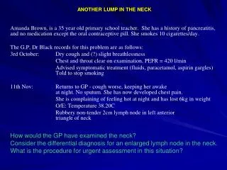
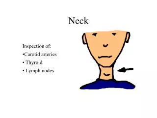
The Case • I saw 8 year old girl brought by mum to A&E at 11pm ,from friend’s home • Mum noticed midline neck swelling 1 week ago, saw GP thought was a lymph node • Friend suggested visiting A&E on eve of presentation as swelling was not settling, infact increasing in size and became red
The Case • Patient was frightened • She had a midline neck swelling with a redness developing at the tip • She was systemically well • She wont cooperate enough to do tongue protrusion • There was no local lymph nodes palpable
Neck Swellings in Children • Neck lumps constitute important diagnostic category • Malignancy less than 1% • Categories: Congenital • Inflammatory/ Infective • Embryological knowledge important for diagnosis and treatment( ?excision)
Branchial cleft apparatus and its derivatives • The branchial arches are ridges, visible in the cervical region of the embryo from the fourth to the eighth week of gestation • 1st arch: mandible, Eustachian tube and some bones of middle ear • 2nd arch: hyoid bone and tonsillarfossa
Branchial derivatives • These may take the form of cysts, sinuses, or cartilaginous remnants, possible to identify the relevant branchial arch from the anatomical position. • Strangely, usually been present since birth, branchial cysts most commonly present in adolescence
Preauricular and first branchial remnants—Small sinuses and cartilage remnants just in front of the ear are the commonest finding but are probably not of branchial origin. • Second branchial remnants—The external opening of a branchial sinus or fistula is almost always related to the anterior border of the sternomastoid
Brachial derivatives • Treatment—Uninfected derivatives should be treated by formal surgical excision, with a careful attempt made to identify any deeper components.
Thyroglossal derivatives • The thyroid gland develops from tissue originally derived from the posterior third of the tongue, which descends during fetal life to its final position anterior to the tracheal rings • Thyroglossalcysts:The key diagnostic features of these neck lumps are their midline position and movement on tongue protrusion and swallowing. Most are intimately related to the hyoid bone, which explains their relation to the tongue and muscles of swallowing.
Thyroglossal cyst-examination and treatment • Although clinical examination is often sufficient for diagnosis, some surgeons obtain a radioisotope thyroid scan before excision to ensure that a normal thyroid gland is present. Excision of the middle third of the hyoid bone in continuity with the cyst (Sistrunk's operation) should be performed to reduce the possibility of recurrence.
Cervicofacialdermoids • The soft tissues of the face are formed by the convergence of three facial processes (frontal, maxillary, and mandibular) • As a consequence, there are lines of fusion where islands of ectodermal tissue may become submerged, later to secrete sebaceous material and present as obvious cystic swellings known as dermoids.
Any suspicion that a dermoid may be fixed to the bone should prompt an x ray examination or even computed tomography to test this possibility. Dermoids should be treated by excision.
Cystic hygroma • These are hamartomatous, lymphatic malformations that result in a multicystic mass which infiltrates tissue planes and has no tendency to spontaneous resolution. Over 60% are found in the neck region, but other sites of origin may include the axilla and chest wall • Treatment: surgical excision or inactivated streptococcal organism-on named patient basis from Japan
Cervical lymphadenopathy, lymphadenitis, and abscess • Characteristic features of lymphadenopathy • Found along jugular vein • Mostly benign • Related to respiratory and throat infections • Histological appearance of reactive hyperplasia
Characteristic features of lymphadenitis • Acute tenderness • Pain • Swelling • Erythema of overlying skin • If pus is formed it requires surgical drainage
Neck Swelllings • Mycobacterial lymphadenitis—If the history of the condition is longer (perhaps over a period of weeks), less acutely tender, and responds only partially or not at all to an appropriate antibiotic then lymphadenitis due to mycobacterial organisms should be considered. In Britain the causative organism is usually an atypical mycobacterium (such as Mycobacterium avium-intracellulare).
Case resolution • Patent disucussed with ENT, BRI asked to prescribe antibiotics • Next day seen there thought was an infected thyroglossal cyst • Plan is after infection settles then surgical excision
Sources • Mainly:ABC of general surgery in children: lumps and swellings of the head and neck M Davenport - BMJ, 1996 - bmj.com • Thanks