Download
1 / 1
50 likes | 442 Vues
Type I Clavicle Fracture: A Case Report Caitlin Ryan, Athletic Training Student. Differential Diagnosis: Dislocation – Acromioclavicular Joint, Glenohumeral Joint. Rib Fractures Pneumothorax Rotator Cuff Pathology Sternoclavicular Joint Injury.

E N D
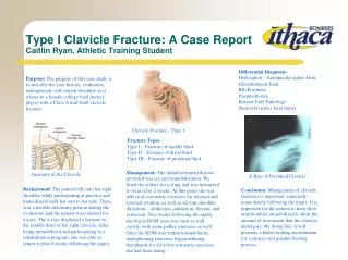
Type I Clavicle Fracture: A Case ReportCaitlin Ryan, Athletic Training Student Differential Diagnosis: Dislocation – Acromioclavicular Joint, Glenohumeral Joint. Rib Fractures Pneumothorax Rotator Cuff Pathology Sternoclavicular Joint Injury Purpose: The purpose of this case study is to describe the case history, evaluation, management, and current literature as it relates to a female college field hockey player with a Class A mid-shaft clavicle fracture. Clavicle Fracture – Type 1 Fracture Types: Type I – Fracture of middle third. Type II – Fracture of distal third. Type III – Fracture of proximal third. Management: The initial treatment that we provided was ice and immobilization. We fitted the athlete for a sling and was instructed to wear it for 2 weeks. At this point she was able to do isometric exercises for internal and external rotation, as well as all four shoulder directions – abduction, adduction, flexion, and extension. Two weeks following the injury, she began ROM exercises such as wall crawls, with some pulley exercises as well. Once the ROM was within normal limits, strengthening exercises began utilizing therabands for all of the isometric exercises she had been doing. Anatomy of the Clavicle X-Ray of Fractured Clavicle Background: The patient fell onto her right shoulder while participating at practice and immediately held her arm to her side. There was a notable deformity present during the evaluation and the patient was referred for x-rays. The x-rays displayed a fracture in the middle third of her right clavicle. After being immobilized and participating in a rehabilitation program, she was able to return to play 6 weeks following the injury. Conclusion: Management of clavicle fractures is important, especially immediately following the injury. It is important for the patient to keep their arm/shoulder immobilized to limit the amount of movement that the clavicle undergoes. By doing this, it will promote a better healing environment for a cleaner and quicker healing process.