Overview of Cellular Structures and Sizes in Microbiology
This document provides a comprehensive overview of the sizes and structures of various cellular entities, from the smallest bacteria measuring at 100 nanometers to larger plant and animal cells observable under light microscopy. It details the dimensions of nerve cells, eggs, and typical bacterial features, such as flagella and cell walls. Additionally, it explains the importance of the surface-to-volume ratio in cellular biology, emphasizing the organization and complexity of both prokaryotic and eukaryotic cells, including their internal components and membrane structures.

Overview of Cellular Structures and Sizes in Microbiology
E N D
Presentation Transcript
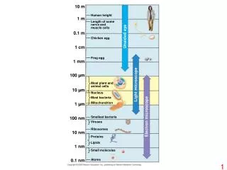
10 m Human height 1 m Length of some nerve and muscle cells 0.1 m Unaided eye Chicken egg 1 cm Frog egg 1 mm 100 µm Most plant and animal cells Light microscope 10 µm Nucleus Most bacteria Mitochondrion 1 µm Electron microscope Smallest bacteria 100 nm Viruses Ribosomes 10 nm Proteins Lipids 1 nm Small molecules Atoms 0.1 nm
Fimbriae Nucleoid Ribosomes Plasma membrane Cell wall Bacterial chromosome Capsule 0.5 µm Flagella (a) A typical rod-shaped bacterium (b) A thin section through the bacterium Bacillus coagulans (TEM)
(a) TEM of a plasma membrane Outside of cell Inside of cell 0.1 µm Carbohydrate side chain Hydrophilic region Hydrophobic region Hydrophilic region Phospholipid Proteins (b) Structure of the plasma membrane
Surface area increases while total volume remains constant 5 1 1 Total surface area [Sum of the surface areas (height width) of all boxes sides number of boxes] 150 750 6 Total volume [height width length number of boxes] 1 125 125 Surface-to-volume (S-to-V) ratio [surface area ÷ volume] 6 1.2 6
Nuclear envelope ENDOPLASMIC RETICULUM (ER) NUCLEUS Nucleolus Rough ER Smooth ER Flagellum Chromatin Centrosome Plasma membrane CYTOSKELETON: Microfilaments Intermediate filaments Microtubules Ribosomes Microvilli Golgi apparatus Peroxisome Mitochondrion Lysosome
Rough endoplasmic reticulum Nuclear envelope Nucleolus NUCLEUS Chromatin Smooth endoplasmic reticulum Ribosomes Central vacuole Golgi apparatus Microfilaments Intermediate filaments CYTO- SKELETON Microtubules Mitochondrion Peroxisome Chloroplast Plasma membrane Cell wall Plasmodesmata Wall of adjacent cell
Central vacuole Cytosol Central vacuole Nucleus Cell wall Chloroplast 5 µm
Nucleus Rough ER Smooth ER cis Golgi Plasma membrane trans Golgi
Intermembrane space Outer membrane Free ribosomes in the mitochondrial matrix Inner membrane Cristae Matrix 0.1 µm
Ribosomes Stroma Inner and outer membranes Granum 1 µm Thylakoid
Direction of swimming (a) Motion of flagella 5 µm Direction of organism’s movement Power stroke Recovery stroke (b) Motion of cilia 15 µm