Download
1 / 28
310 likes | 383 Vues
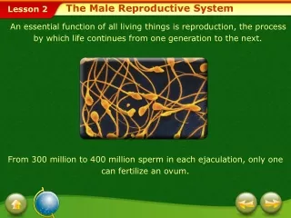
Explore the development and anatomy of the male reproductive system, from descent of the testes to sperm production and transport. Learn about accessory glands and histology.

E N D
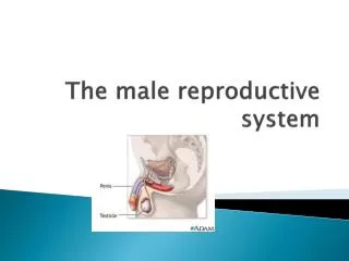
The Male Reproductive System Exercise 30 A&P 233
The Reproductive System • Male and female reproductive systems develop from similar embryonic tissue. • First few weeks of development, male and female embryos are indistinguishable. • Adult reproductive systems share some functional similarities.
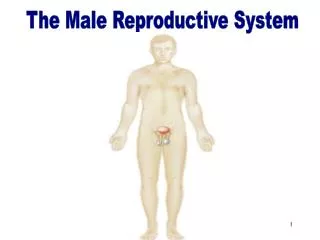
Descent of the Testes • Ovoid structures about 5 cm long and 3 cm wide. • Located within the scrotal sac (scrotum) • During fetal development they are near the kidneys and slowly move inferiorly in the abdominal cavity. • During the 7th month they descend through the inguinal canals
Scrotum • Sac of skin and superficial fascia that hangs outside the abdominopelvic cavity at the root of the penis • Contains paired testicles separated by a midline septum • Its external positioning keeps the testes 3C lower than core body temperature
Wall of the Scrotum • In the dermis, there is a thin layer of smooth muscle known as the dartos muscle. Contractions of this muscle causes wrinkling of the skin. • The cremaster muscle is a thicker layer of skeletal muscle that lowers and raises the testes based on temperature.
Inside the Scrotum • Each testes is enclosed by the tunicavaginalis, a continuation of the peritoneum that lines the abdominopelvic cavity. • A fibrous capsule covers each testis called the tunicaalbuginea.
Testicle • The tunica albuginea gives rise to septa (partitions) that divide the testis into lobules (about 250) • Each lobule contains 3 or 4 highly coiled seminiferous tubules • These converge to become rete testis which transport sperm to the epididymis
Spermatic Cord • Contains the structures running from the testicles to the pelvic cavity. • Passes through the inguinal canal • Contents: • Vas Deferens • Nerves • Blood Vessels
Accessory Glands: Seminal Vesicles • Lie on the posterior wall of the bladder and secrete 60% of the volume of semen • Seminal fluid: • Fructose: provides energy for the sperm. • Fibrinogen: helps turn semen into a bolus that can be readily propelled into the vagina. • Prostaglandins: decrease cervical mucus viscosity and stimulate reverse peristalsis of the uterus. • Join the ductus deferens to form the ejaculatory duct
Accessory Glands: Prostate Gland • Doughnut-shaped gland that encircles part of the urethra inferior to the bladder • Plays a role in the activation of sperm • Enters the prostatic urethra during ejaculation • Prostatic secretions include: • Citrate: is a food source (TCA cycle) • Proteolytic enzymes: acts to "decoagulate" the semen that was coagulated by seminal vesicle secretions, which helps the sperm begin their journey once inside the vagina
Bulbourethral Glands (Cowper’s Glands) • Pea-sized glands inferior to the prostate • Produce alkaline mucus prior to ejaculation that neutralizes traces of acidic urine in the urethra
Epididymis • Epididymis: Storage and maturation area for sperm • Its head joins the efferent ductules and caps the superior aspect of the testis • The duct of the epididymis has stereocilia that: • Absorb testicular fluid • Pass nutrients to the sperm • Nonmotile sperm enter, pass through its tubes and become motile (propelled by peristalsis) • Upon ejaculation the epididymis contracts, expelling sperm into the ductus deferens
Review questions • What is the difference between mitosis and meiosis? • What are the results of meiosis?
Spermatogenesis • Spermatogenic stem cells of the seminiferous tubules give rise to sperm in a series of events • Mitosis of spermatogonia, forming spermatocytes • Meiosis forms spermatids from spermatocytes • Spermiogenesis: spermatids form sperm
Sperm • Sperm have three major regions • Head :contains DNA and has a helmet-like acrosome containing hydrolytic enzymes that allow the sperm to penetrate and enter the egg • Midpiece: contains mitochondria spiraled around the tail filaments • Tail :a typical flagellum produced by a centriole
Sperm Summary • Produced: Seminiferous tubules • Stored: Epididymis • Transported through epididymis by rhythmic peristaltic contractions as they mature • EpididymisVas DeferensEjaculatory duct (ampulla of vas deferens fuses with duct of seminal vesicle “ejaculatory duct”) prostate prostatic urethra (then passes the bulbourethral gland) membranous urethrapenile urethra
Today’s lab • ID structures on the models • View slides of testes, penis, sperm