Overview of Cardiovascular Health: Blood Counts, Vascular System, and Cardiac Function
380 likes | 485 Vues
This overview discusses various aspects of cardiovascular health, including abnormal blood counts such as polycythemia vera and anemia, and their implications for diseases like leukemia and AIDS. It covers the fluid spaces in the body, arterial and venous structures, and the critical role of the vascular system. The guide also explains the significance of heart sounds, electrocardiogram (ECG) findings, electrolyte imbalances, and conditions such as mitral valve prolapse. Understanding these components is essential for maintaining cardiovascular health.

Overview of Cardiovascular Health: Blood Counts, Vascular System, and Cardiac Function
E N D
Presentation Transcript
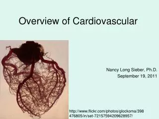
Overview of Cardiovascular Nancy Long Sieber, Ph.D. September 19, 2011 http://www.flickr.com/photos/glockoma/398476805/in/set-72157594209628957/
Abnormal Blood Counts • Too many red blood cells and platelets: polycythemia vera, certain blood cancers • Too few red blood cells: anemia, due to insuffient iron in the diet, hemolysis, chemotherapy • Too many white blood cells: leukemia and other other blood cancers • Too few white blood cells: AIDS, other immune deficiencies, chemotherapy
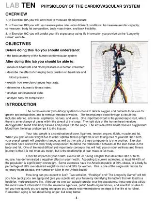
Fluid Spaces • Intravascular (first space) • Interstitial (second space) • “Third space” - interstitial spaces that expand as the result of injury or disease. Eg: swelling at site of surgical incision, edema in abdominal cavity.
Elephantiasis: The swelling results from blocked lymphatic vessels
The distribution of blood flow at rest and during exercise. Note how blood conditioning organs (the kidneys and abdominal organs) are able to withstand a significant reduction in blood flow during exercise.
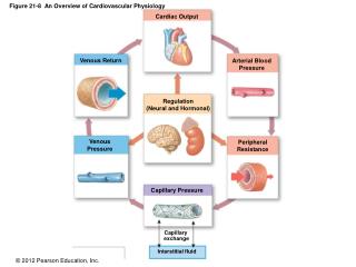
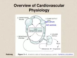
Arteries Low resistance conduits Highly elastic Arterioles Major site where total peripheral resistance (TPR) is controlled Control of blood flow to organs and tissues Capillaries and venules Exchange of nutrients, waste, and fluid between the blood and interstitium Largest cross-sectional area and slowest flow Veins Low resistance conduits that return blood to the heart Blood storage reservoir – can be mobilized as needed The vascular system
An Aortic Aneurysm http://www.nlm.nih.gov/medlineplus/ency/imagepages/18072.htm
Varicose Veins http://www.vascularweb.org/graphics/northpoint_graphics_jpg/Varicose_02_Base_300.jpg
Cardiac Muscle http://content.answers.com/main/content/img/oxford/Oxford_ Body/019852403x.cardiac-muscle.1.jpg
Atrial Fibrillation http://www.lhsc.on.ca/uwodoc/media/fib2.gif
Two normal beats, followed by ventricular fibrillation http://noodle.med.yale.edu/~staib/bme355/ecg/vfib.jpg
Hypothermia can lead to a characteristic Osborn (or “J”) wave at the end of the QRS complex. A 12-lead ECG obtained at core body temperature of 85°F. Note Osborn waves, an extra deflection at end of QRS complex (arrows). From:Circulation June 27, 2000 vol. 101 no. 25 e233-e244
Electrolyte imbalances can interfere with the cardiac rhythm, potentially leading to cardiac arrest. Examples: • Eating disorders and other types of malnutrition. • Hyponatremia – can result from over consumption of water. • People who are on antidiuretic drugs may also develop levels of K+ that are too high or too low.
Heart Sounds http://www.nlm.nih.gov/medlineplus/ency/images/ency/fullsize/19613.jpg
Heart SoundsS1 and S2 are normal S3 and S4 are abnormal http://connection.lww.com/Products/taylor5e/documents/Ch25/jpg/25_039.jpg
Extra Diastolic Sounds: S3 • S3 is produced by the tensing of the chordae tendineae, which occurs during rapid filling and expansion of the ventricles. • Common in children and young adults – the flexible ventricles of young people can expand rapidly. • In middle-aged or older adults often indicates excessive volume in the ventricles, which usually indicates heart failure. www.mvprolapse.com/mvp.html
Extra Diastolic Sounds: S4 • Produced by the left or right venticle contracting against a stiffened ventricle • Usually indicates a loss of compliance of the ventricle due to ventricular hypertrophy or myocardial ischemia
The heart murmur associated with mitral valve prolapse Mitral valve prolapse causes a late systolic murmur
Mitral Valve Prolapse Mitral valve prolapse is an example of valve insufficiency. Abnormally shaped valve leaflets are pushed into the left atrium during late systole. http://www.nlm.nih.gov/medlineplus/ency/images/ency/fullsize/18148.jpg
The length-tension relationship http://www.abcbodybuilding.com/magazine03/wrench/tensionlevels2.jpg
Children with Kwashiorkor http://www.cs.stedwards.edu/chem/Chemistry/CHEM43/CHEM43/Leukotr/Kwashiorkor.GIF