Introduction to Neuroanatomy
Introduction to Neuroanatomy. Russell M. Bauer, Ph.D. January 9, 2006. Caveats. What you need to know about anatomy Changes with each individual case Involves both local and global knowledge Depends on your practice and referral question What I can give you in 90 minutes

Introduction to Neuroanatomy
E N D
Presentation Transcript
Introduction to Neuroanatomy Russell M. Bauer, Ph.D. January 9, 2006
Caveats • What you need to know about anatomy • Changes with each individual case • Involves both local and global knowledge • Depends on your practice and referral question • What I can give you in 90 minutes • Conceptual understanding of organization • Tools to think about deeper levels of analysis • Ways of decomposing deficits
Key Concepts • Functional Systems: patterns of connectivity • Localized damage has systemic effects • Segregated patterns of inputs and outputs • Excitatory and inhibitory control • Parallel vs. serial processing
Directions and Planes of Section Horizontal (Axial) Coronal Sagittal
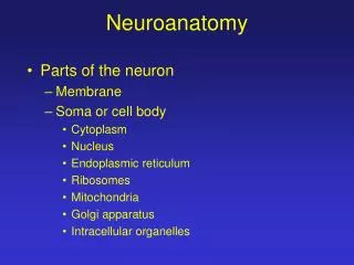
Cerebral Cortex • The layer of gray matter covering • the entire surface of cerebral hemisphere • Migration of neurons from inner mantle layer of • neural tube • Accommodates enormous number of neurons • -Large surface areaaccommodates • more neurons than deep nuclei • -Gyri and sulcialso increase surface area • -Laminar organizationalso accommodates • enormous number of neurons Won Taek Lee, M.D., Ph.D.
CerebralCortex Numerical Data Total surface area: 2200 cm2 (2.5 ft2) about 1/3 ------ surface area about 2/3 ------ hidden in the sulci Thickness: 1.5 mm (V I) - 4.5 mm (M I) Generally, thickest over the crest of the convolution and, thinnest in the depth of sulci Weight: 600 gm (40 % of total brain weight) 180 gm --------- neurons 420 gm --------- glial cells Won Taek Lee, M.D., Ph.D.
CerebralCortex Numerical Data Number of neuronal cells in cerebral cortex neurons ----------- 10-15 billion glial cells ---------- 50 billion Estimation of number of cortical neurons von Economo and Koskinas (1925) 14.0 billion Shariff (1953) 6.9 billion Sholl (1956) 5.0 billion Pakkenberg (1966) 2.6 billion Won Taek Lee, M.D., Ph.D.
Subdivision of Cerebral Cortex Allocortex Archicortex (hippocampal formation) Palaeocortex (Paleopallium) Isocortex Neocortex (Neopallium) Won Taek Lee, M.D., Ph.D.
Isocortex– typical 6 layered cortex I. Molecular Layer II. External Granular Layer III. External Pyramidal Layer IV. Internal Granular Layer V. Internal Pyramidal Layer VI. Polymorphic Layer Won Taek Lee, M.D., Ph.D.
CerebralCortex 1. Pyramidal Cell 2. Fusiform Cell 3. Granular (Stellate) Cell 4. basket cell 5. double bouquet cell 6. chandlier cell 7. neurogliform cell 8. Horizontal Cell of Cajal 9. Cells of Martinotti a: axon Won Taek Lee, M.D., Ph.D.
Columnar Cortical Unit and Cortical Circuitry A. pyramidal neuron B. excitatory granular cell C.inhibitory granular cell 1. afferent fiber 2.efferent fiber 3.corticothalamic fiber Won Taek Lee, M.D., Ph.D.
1 3 3 2 1 2 1 3 Order of Cortical Maturation
Somesthetic Area S I (3, 1, 2 ; postcentral gyrus) afferents: ventrobasal complex (VPLc, VPM) discrimination of position and intensity of sensation S II (superior bank of lateral fissure) Somesthetic Association Cortex (5, 7; parietal lobe, precuneus) afferents: S I, LP of thalamus integration of general sensation with past experience tactile agnosia, astereognosis
Primary Somesthetic Area Thalmocortical connection (VPLc S I) Central region --- cutaneous (3b, 1) Peripheral region --- deep (3a, 2)
Secondary Somesthetic Area (SII) superior bank of lateral fissure
Kaas JH & Catania, KC (2002). How do features of sensory representations develop? Bioessays, 24, 334-343.
Motor Areas primary Motor Area (M I) Premotor Area (PM) Supplementary Motor Area (SMA) Frontal Eye Field
Association Areas Language Areas: 22, 39, 40, 44, 45 Posterior Parietal Association Area: 5, 7(39, 40) body image Temporal Association Area: 20, 21, 37, 38 (22) multisensory integration, conceptual ideation Prefrontal Association Area: 9, 10, 11, 12, 46, 47 (44,45) executive skills, judgment, planning, emotion-regulation
Neuropsychological Manifestations of Temporal Lobe Lesions Lateral (20,21,38,37) A) Posterior: visual recognition disturb-ances (severity depends on whether unilateral or bilateral B) Anterior: anomia for objects (left), anomia for facial expressions (right), full-blown anomia, retrograde memory disturbance (bilateral) Mesial (27,28 and limbic structures) Anterograde amnesia for verbal (left) or nonverbal (right) material; bilateral lesions produce severe material non-specific defect Tranel, 1992
Neuropsychological Manifestations of Occipital Lobe Lesions Dorsal(17,18,19,39,7) Partial or mild Balint’s syndrome (unilateral), severe Balint’s syndrome (bilateral), defective motion perception, astereopsis, severe visuospatial disturbance, simultanagnosia Ventral (17,18,19) Hemiachromatopsia (unilateral), pure alexia (left), apperceptive visual agnosia (unilateral, R>L), defective imagery Bilateral lesions produce severe agnosia, prosopagnosia Bilateral lesions of Area 17 produce cortical blindness (may be transient or evolving) Tranel, 1992
Neuropsychological Manifestations of Parietal Lobe Lesions Temporoparietal Junction (including posterior superior temporal gyrus, 22, inferior 39,40) A)Left: Wernicke’s aphasia B) Right: Amusia, defective music recognition, ‘phonagnosia’ C) Bilateral: auditory agnosia Inferior Parietal Lobule (39,40) A) Left: Conduction aphasia, tactile agnosia B) Right: neglect, anosognosia, tactile agnosia, anosodiaphoria C) Balint’s syndrome Tranel, 1992
Neuropsychological Manifestations of Frontal Lesions I Frontal Operculum (44,45,47) A) Left: Broca’s aphasia B) Right: ‘expressive’ aprosodia Superior Mesial (mesial 6, 24) A)Left: akinetic mutism B) Right: akinetic mutism Bilateral lesions of mesial SMA (6) and anterior cingulate (24) produce more severe form of akinetic mutism Tranel, 1992
Neuropsychological Manifestations of Frontal Lobe Lesions II Inferior Mesial Region A)Orbital Region (10, 11) Lesions in this region produce disinhibition, altered social conduct, “acquired sociopathy”, and other disturbances due to impairment in fronto-limbic relationships B)Basal Forebrain (posterior extension of inferomesial region, including diagonal band of Broca, nucleus accumbens, septal nuclei, substantia innominata Lesions here produce prominent anterograde amnesia with confabulation (material specificity present, but relatively weak) Tranel, 1992
Neuropsychological Manifestations of Frontal Lobe Lesions III Lateral Prefrontal Region (8,9,46) Lesions in this region produce impairment in a variety of “executive” skills that cut across domains. Some degree of material-specificity is present, but relatively weak. A) Fluency: impaired verbal fluency (left) or design fluency (right) B) Memory impairments: defective recency judgment, metamemory defects, difficulties in memory monitoring C) Impaired abstract concept formation and hypothesis testing D) Defective planning, motor sequencing E) Defective cognitive judgement and estimation Tranel, 1992
Blood Supply to Cortical and Deep Structures Blumenfeld, 2002