Download
1 / 29
310 likes | 790 Vues
Learn the physical examination techniques for assessing chest and lung health, including inspection, palpation, percussion, and auscultation. Master the skills that evolve over time with practice. Identify crucial signs during head and neck, eyes, and lung topography examinations for a detailed clinical evaluation.

E N D
Physical examination employs the use of inspection, palpation, percussion, and auscultation to determine patients’ clinical status and their response to therapy • Each examination is modified according to the purpose of the examination • Physical examination skills develop over time with practice
Examination of the Headand Neck • Identify the patient’s facial expression, looking for evidence of pain or acute distress • Look for evidence of cyanosis around the lips and oral mucosa • Patients may use pursed-lip breathing when COPD is present
Eyes • The eyes are inspected for pupillary response to light when neurologic defects are suspected • Dilated and fixed pupils suggest brain death in some patients • The eyelids may droop (ptosis), indicating damage to the third cranial nerve
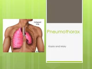
Neck • The trachea should be midline • If it is deviated to one side, a unilateral lung problem is probably present • Atelectasis • pneumothorax • The status of the jugular veins in the neck is important • Patients with cor pulmonale have JVD • Use of accessory muscles in the neck suggests obstructive lung disease
Lung Topography • Anterior chest is defined by the midsternal and midclavicular lines
Lung Topography • Lateral chest is defined by midaxillary, anterior axillary and posterior axillary lines
Lung Topography • Posterior chest is defined by the midspinal and midscapular lines
Thoracic Cage Landmarks • On the posterior chest, C7 is seen as the most prominent spinous process at the base of the neck
Thoracic Cage Landmarks • The angle of Louis, or sternal angle, is located on the anterior chest. • Formed by the ridge between the manubrium and the gladiolus
Lung Fissures • The oblique fissure • starts at rib six on the anterior chest at the midclavicular line • It runs up and laterally crosses the midaxillary line at fifth rib and across the posterior chest, ending at T3 • horizontal fissure • passes from the fourth rib at the midsternal line laterally to the fifth rib in the midaxillary line
Tracheal Bifurcation • At T4 on posterior chest • At sternal angle on anterior chest
Diaphragm • The diaphragm is a dome-shaped muscle • The top of the dome rests at about the fifth rib anteriorly and at T9 on the posterior chest normally
Lung Borders • On the anterior chest the upper border of the lung extends 2 to 4 cm above the medial third of the clavicles. The inferior border of the lung is at rib six normally
Lung Borders • On the lateral chest the lower margin of the lung is at rib eight
Lung Borders • On the posterior chest the superior border of the lung extends to T1. The inferior border varies with breathing but is usually at about T10
Examination of the Thorax • Look • Feel • Listen
Look For • A barrel chest or evaluate the A-P diameter • An in crease A-P diameter is consistent with COPD
Look For • Kyphoscoliosis is present when the spine is bent laterally and from front to back • Can causea restrictive lung problem
Look For • Pectus carinatum is seen as an abnormal sternal protrusion
Look For • Pectus excavatum is seen as depression of the sternum
Look For • Breathing pattern is important to identify when lung disease is present • Rapid and shallow breathing is consistent with restrictive disease • A prolonged expiratory time is consistent with obstructive lung disease • Retractions are seen as inward depression of the skin around the rib cage with inspiration • This suggests a high work of breathing (WOB) • Abdominal paradox is seen as inward movement of the abdomen with inspiration • This suggests diaphragm paralysis or fatigue • Hoover’s sign is seen as inward movement of the lateral chest with inspiration. It is a sign of severe COPD.
Feel For (Palpation) • Vocal fremitus is assessed to identify pathologic changes in the lung. • Increased vocal fremitus is consistent with pneumonia and atelectasis. • Decreased vocal fremitus is consistent with lung hyperinflation, pleural disorders, and obesity.
Palpation • Use palpation to assess for uniform chest excursion
Percussion • Percussion is done to determine the condition of the underlying lung. • Increased resonance is heard with pneumothorax and lung hyperinflation. • Decreased resonance is heard with pneumonia and atelectasis.