Download
1 / 34
370 likes | 1.71k Vues
Chest Wall, Lung, Mediastinum, & Pleura. May 11, 2010. Trachea. Tracheal Injury. Tracheal Stenosis Over-inflation of the cuff Ischemia, scarring, stricture Fistula development Incorrect placement of the tracheostomy through the first tracheal ring or the cricothyroid membrane

E N D
Chest Wall, Lung, Mediastinum, & Pleura May 11, 2010
Tracheal Injury • Tracheal Stenosis • Over-inflation of the cuff • Ischemia, scarring, stricture • Fistula development • Incorrect placement of the tracheostomy through the first tracheal ring or the cricothyroid membrane • Use of a large tracheostomy tube • Stridor and dyspnea on exertion • Resection and primary anastomosis
TracheoInnominate Artery Fistula • Causes • Too low placement of the tracheostomy (below the 4th ring) • Hyperinflation of the tracheal cuff • Typically occur 2 weeks after tracheostomy • Signs • Sentinel bleed • Heavy bleed – blow up cuff • Insert finger and press against manubrium • Oral intubation and emergent resection of fistlua
TracheoEsophageal Fistula • Causes • ETT cuff compresses against NG tube • Signs • Gastric contents or tube feeds suctioned from airway • Gastric distention from positive pressure ventilation • Diagnosis • Bronchoscopy or EGD • Treatment • Wean off vent • Remove NG and place GT or JT • Operative repair involves resection and primary repair of tracheal pathology, repair of esophagus, and interposition muscle flap
Tracheal Neoplasms • Rare • SCC or adenoid cystic carcinoma • Cough, dyspnea, hemoptysis, stridor, or symptoms of invasion of contiguous structures • 50% of patients have tracheal stenosis on Xray • 50% have stage IV dz at time of diagnosis • Overall 5-year survival is 40%, but falls to 15% for those with stage IV disease
Surgical Approaches to the Thoracic Cavity • Posterior Thoracotomy – most common approach for pulmonary resections, esophageal procedures, posterior mediastinal access, and vertebral procedures • Patient in lateral decubitus – risk of injury to brachial plexus or axillary vascular structure • Anteriolateral Thoracotomy – trauma victims • Median sternotomy – cardiac procedures • VATS – improved pain and functional recovery • Improved ability to tolerate chemotherapy • Quicker return of respiratory function in elderly and COPD
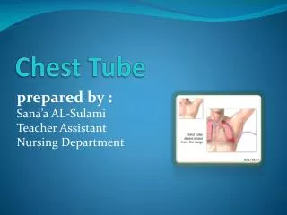
Post-operative Care • Chest Tube • Evacuation of air • Evacuation of blood/pleural fluid • Check the system regularly • Pain Control • Epidural at T6 with ropivicaine • Hypotension and urinary retention • Narcotics and Toradol • Aggressive Pulmonary Toilet
Post-operative Complications • Air leak • Bronchopleural fistula • Diagnose by bronchoscopy • Continued chest tube mgmt • Operative closure with intercostal muscle flap • Bronchoscopic fibrin glue application (<4mm) • Concomitant empyema may require open drainage • Post-pneumonectomy pulmonary edema • Decreased lympnatic drainage • Mechanical ventilation & diuresis
Lung • Solitary Pulmonary Nodule • A single, well-circumscribed, spherical lesion • ≤3 cm in diameter • Completely surrounded by normal aerated lung parenchyma • Detected incidentally on chest radiographs or CT scans • DDx – malignancy, hamartoma (10%), granulomatous dz (70%) • 20 to 40% likelihood of being malignant • 50% or higher in smokers • growth over time, • density of the lesion on CT • associated symptom • age, sex, cigarette smoking history, occupational history, • prevalence of endemic granulomatous disease
Lung Cancer • Leading cause of cancer-related death (30%) • Second most common diagnosed cancer • Women – breast • Men – prostate
Lung Cancer • Epidemiology • Leading risk factor – smoking (polycyclic aromatic hydrocarbons) • Secondhand smoke also increases risk • Environmental exposures - asbestos, arsenic, and chromium compounds • COPD • h/o tuberculosis
Lung Cancer • Non-small cell carcinoma vs neuroendocrine tumors • NSCLC - large cell carcinoma, squamous cell carcinoma, adenocarcinoma, and BAC • Clinical behavior and treatment options are similar and thought of as a uniform group • Neuroendocrine - typical carcinoid, atypical carcinoid, large cell neuroendocrine carcinoma, and small cell carcinoma
Squamous Cell Carcinoma • 30 to 40% of lung cancers • Highly associated with cigarette smoking • Primarily located centrally and arises in the major bronchi • Typical symptoms are hemoptysis, bronchial obstruction with atelectasis, dyspnea, and pneumonia • Peripherally based SCC will develop in a tuberculosis scar or in the wall of a bronchiectatic cavity • Central necrosis is frequent and may lead to the radiographic findings of a cavity (air-fluid level) • May become infected & form an abscess
Adenocarcinoma • Incidence has increased over the last several decades & now 25 to 40% of all lung cancers. • Most frequent histologic type found in women • Peripherally based tumor • Frequently discovered incidentally on routine chest radiographs • Symptoms of chest wall invasion or malignant pleural effusions • Composed of glands with or without mucin production and destruction of contiguous lung architecture
Bronchoalveolar Carcinoma • Unusual (5% of all lung cancers) subtype of adenocarcinoma • Unique growth pattern • Rather than invading and destroying contiguous lung parenchyma, tumor cells multiply and fill the alveolar spaces • Because of their growth within alveoli, BAC tumor cells from one site can aerogenously seed other parts of the same lobe or lung, or the contralateral lung. • Three radiographic presentations: • a single nodule • multiple nodules (in single or multiple lobes) • diffuse form with an appearance mimicking that of a lobar pneumonia • Air bronchograms can be seen
Large Cell Carcinoma • 10 to 20% of lung cancers and may be located centrally or peripherally • Often admixed with other cell types such as squamous cells or adenocarcinoma • May be confused with a large cell variant of neuroendocrine carcinoma, with immunohistochemical staining usually allowing diagnostic distinction between the two
Neuroendocrine Carcinoma • Grade I NEC (classic or typical carcinoid) is a low-grade NEC • primarily in the central airways • primarily in younger patients. • classically presents with hemoptysis, with or without airway obstruction and pneumonia • Regional lymph node metastases are seen in 15% of patients but rarely spread systemically or cause death • Grade II NEC (atypical carcinoid) tumors with a degree of aggressive clinical behavior • linked to cigarette & peripherally located • Much higher malignant potential. • Lymph node metastases found in 30 to 50% of patients • At the time of diagnosis, 25% have remote metastases • Grade III NEC large cell–type tumors occur primarily in heavy smokers • Grade III NEC small cell type [small cell lung carcinoma (SCLC)] is the most malignant NEC and accounts for 25% of all lung cancers • Immunohistochemical stains distinguish from NSCLC • Leading producer of paraneoplastic syndromes
Paraneoplastic Syndromes • Table 19-5 • Hypercalcemia (ectopic parathyroid hormone) • Cushing's syndrome • Syndrome of inappropriate secretion of antidiuretic hormone • Carcinoid syndrome • Gynecomastia • Hypercalcitoninemia • Elevated growth hormone level • Elevated levels of prolactin, follicle-stimulating hormone, luteinizing hormone • Hypoglycemia • Hyperthyroidism • Neuropathy
Metastatic Symptoms • Lung cancer metastases occur most commonly to the CNS, vertebral bodies, bone, liver, adrenal glands, lungs, skin, and soft tissue • At diagnosis, 10% of patients have CNS metastases • another 10 to 15% will go on to develop CNS metastases after diagnosis. • Focal symptoms are most common and include headache, nausea and vomiting, seizures, hemiplegia, and speech difficulty • Most common cause of spinal cord compression • Invasion of an intervertebral foramen from a primary tumor contiguous with the spine • Direct extension of a vertebral metastasis. • Bony metastases, are identified in 25% - lytic and painful • Liver & adrenal metastases are typically asymptomatic and discovered by routine CT scan • Skin and soft tissue metastases occur in 8% of patients dying of lung cancer and generally present as painless subcutaneous or intramuscular masses
Assessment of Primary Lung Cancer • History and directed questions regarding the presence or absence of pulmonary, nonpulmonary, thoracic, and paraneoplastic symptoms • Imaging • Nodes and invasion • Tissue diagnosis • Bronchoscopy or percutaneous biopsy • Thoracoscopy or rarely thoracotomy
Staging • Mediastinal nodes • Stations 4 & 7 • CT scan & perc biopsy • PET • Bronchoscopy • Mediastinoscopy • EUS w FNA • Distant mets • PET-CT
Mediastinal Tumors • Anterior (thymus) • Thymoma – always resect • 50% malignant • 50% have symptoms • 50% have myasthenia gravis • Thyroid cancer and goiter • T-cell lymphoma – treat with XRT and chemo • Teratoma – resection and chemo • Seminoma – resectiona and XRT • Parathyroid adenoma
Mediastinal Tumors • Middle (heart, trachea, ascending aorta) • Bronchiogenic cyst – posterior to the carina - resect • Pericardial cyst – at right costophrenic angle – resect • Enteric cyst - resect • Lymphoma • Posterior (esophagus, descending aorta) • Enteric cysts • Neurogenic tumors – cause pain and neurologic deficit – resect – 10% have intraspinal involvement • Lymphoma
Pleural Disease • Pleural effusion - any significant collection of fluid within the pleural space • Transudative vs exudative • Chylothorax – injury to thoracic duct
Pleural Tumors • Malignant mesothelioma • 50% associated with asbestos exposure • Patients present with dyspnea and chest pain • Treatment options include supportive care only, surgical resection, and multimodality approaches • Fibrous Tumors • Unrelated to asbestos exposure or malignant mesothelioma • Single pedunculated mass arising from the visceral pleura • Found incidnetally • Benign or malignant • Symptoms such as cough, chest pain, and dyspnea occur in 30 to 40% of patients • Less common are fever, hypertrophic pulmonary osteoarthropathy, hemoptysis, and hypoglycemia • Cured by complete surgical resection