Download
1 / 37
930 likes | 3.44k Vues
Imaging Anatomy of the Mediastinum. Dr. Flip Otto Dept. of Radiology Universitas Academic Hospital. Outline. Mediastinal devisions Content of mediastinum Mediastinal contours on PA chest radiograph Cross sectional anatomy of mediastinum

E N D
Imaging Anatomy of the Mediastinum Dr. Flip Otto Dept. of Radiology Universitas Academic Hospital
Outline • Mediastinaldevisions • Content of mediastinum • Mediastinal contours on PA chest radiograph • Cross sectional anatomy of mediastinum • Mediastinal lines and stripes on conventional radiography and CT correlation • Mediastinal spaces • Mediastinallymphnodes
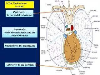
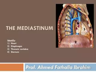
Mediastinaldevisions Devisions used to describe location of pathological processes: • Superior mediastinum: • Above line from lower border T4 to sternal angle • Anterior mediastinum: • Between anterior part of heart and sternum • Middle mediastinum: • Occupied by heart and its vessels • Posterior mediastinum: • Between posterior part of heart and thoracic spine
Cross sectional Anatomy on CTT3 level 4 3 5 6 7 1 2 1-trachea 2-oesophagus 3-right braciocephalic vein 4-left brachiocephalic vein 5-right brachiocephalic artery 6-left common carotid artery 7-left subclavian artery
Cross sectional Anatomy on CTT4 level 4 1 2 5 3 1-trachea 2-aortic arch 3-oesophagus 4-superior vena cava 5-arch of azygos vein
Cross sectional Anatomy on CTT5 level 3 1 7 11 2 6 5 8 12 10 4 9 1-main pulmonary trunk 2-right pulmonary artery 3-ascending aorta 4-descending aorta 5-left main bronchus 6-right main bronchus 7-superior vena cava 8-oesophagus 9-azygos vein 10-azygoesophageal recess 11-left superior pulmonary vein 12-left descending lower-lobe artery
Cross sectional Anatomy on CTT6 level 2 3 1 4 6 5 7 1-aortic root 2-right ventricular outflow tract 3-right atrial appendage 4-left atrium 5-descending aorta 6-oesophagus 7-azygos vein
Cross sectional Anatomy on CTT8 level 2 1 5 4 3 7 6 8 1-right atrium 2-right ventricle 3-left atrium 4-left ventricle 5-left ventricular outflow tract and aortic valve 6-descending aorta 7-oesophagus 8-azygos vein
Cross sectional Anatomy on CTT10 level 3 4 2 1 5 1-descending aorta 2-fundus of stomach 3-inferior vena cava 4-oesophago-gastric junction 5-spleen
Mediastinal lines and stripes- Correlation between plain chest radiography and CT • Lines: • Anterior junction line • Posterior junction line • Stripes: • Right paratracheal stripe • Left paratracheal stripe • Posterior tracheal stripe • Posterior wall of bronchus intermedius • Interfaces: • Right paraspinal line • Left paraspinal line • Aortic-pulmonary stripe • Azygo-oesophageal recess
Abnormal anterior junction line Volume loss in right lung with rightward displacement of anterior junction line following a right middle lobectomy
Abnormal right paratracheal stripe Widening of right paratracheal stripe caused by a large ectopic parathyroid adenoma. Note diffuse osteopenia from hyperparathyroidism.
Abnormal left paratracheal stripe Widening of left paratracheal stripe with mass effect on the trachea due to large thyroid carcinoma and associated supraclavicular lymphadenopathy
Abnormal posterior tracheal stripe Widening of the posterior tracheal stripe due to dilated esophagus in a patient with achalasia.
Abnormal posterior wall of bronchus intermedius Diffuse bandlike thickening of the posterior wall of the bronchus intermedius in a patient with pulmonary oedema.
Abnormal right paraspinal line Abnormal bulge in right paraspinal line inferiorly due to mediastinal hematoma from multiple right sided transverse process fractures and an associated hemothorax.
Abnormal left paraspinal line Focal lateral bulge in left paraspinal line due to extensive esophagealvarices in patient with liver cirrhosis.
Abnormal aortic-pulmonary stripe Abnormal contour of the aortic-pulmonary stripe due to lymphoma with anterior mediastinal lymphadenopathy within the prevascular space.
Abnormal azygoesophageal recess Abnormal contour and right lateral convexity of distal third of azygoesophageal recess due to a large hiatal hernia.
Mediastinal spaces • Four named spaces surrounding the central airways: • Pretracheal space • Aortopulmonary window • Subcarinal space • Right paratracheal space
Abnormal aortopulmonary window Abnormal bulge in AP window due to significant soft tissue mass within AP window and subcarinal space compatible with metastatic lymphadenopathy in a patient with bronchogenic carcinoma. Also widened right paratracheal stripe due to lymphadenopathy and left lower lobe consolidation.
Mediastinallymphnodes • American Thoracic Society definitions of regional lymph node stations • X Supraclavicular nodes • 2R Right upper paratracheal nodes • 2L Left upper paratracheal nodes • 4R Right lower paratracheal nodes • 4L Left lower paratracheal nodes • 5 Aortopulmonary nodes • 6 Anterior mediastinal nodes • 7 Subcarinal nodes • 8 Paraesophageal nodes • 9 Right or left pulmonary ligament nodes • 10R Right tracheobronchial nodes • 10L Left tracheobroncheal nodes • 11 Intrapulmonary nodes
Conclusion • Traditional frontal and lateral chest radiography remains a valuable tool in the evaluation of chest disease despite increased reliance on CT, therefore familiarity with anatomic basis of mediastinal lines and stripes as seen on radiography imperative. • Knowledge of normal anatomic structures within different mediastinal divisions helps guide formulation of appropriate differential diagnosis
References • Butler, P., Mitchell, A.W.M., Ellis, H. (1999). Applied Radiological Anatomy. Cambridge: Cambridge University Press • Ellis, H., Logan, B.M., Dixon, A.K. (2007). Human Sectional Anatomy – Atlas of body sections, CT and MRI images, 3rd ed. London: Hodder Arnold • Gibbs, J.M., Chandrasekhar, C.A., Ferguson, E.C. et al. (2007). Lines and Stripes: Where did they go? – From Conventional Radiography to CT. Radiographics, 27:33-48. • Netter, F.H. (2011). Atlas of Human Anatomy, 5th ed. Philadelphia: Saunders Elsevier • Ryan, S., McNicholas, M., Eustace, S. (2011). Anatomy for diagnostic imaging, 3rd ed. London: Saunders Elsevier