THE MIDDLE MEDIASTINUM
350 likes | 564 Vues
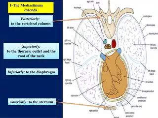
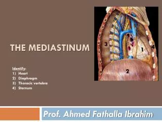
THE MIDDLE MEDIASTINUM. Middle mediastinum. C ontains. THE Pericardial Sac. INCLUDES. Heart. Origins of the great vessels: Ascending Aorta Pulmonary trunk Lower half of superior vena cava Small part of inferior vena cava very small part of Pulmonary veins. Ascending Aorta.

THE MIDDLE MEDIASTINUM
E N D
Presentation Transcript
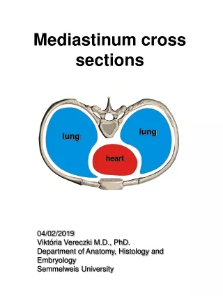
Middle mediastinum Contains THE Pericardial Sac INCLUDES Heart Origins of the great vessels: Ascending Aorta Pulmonary trunk Lower half of superior vena cava Small part of inferior vena cava very small part of Pulmonary veins
Ascending Aorta • The ascending aorta lies within the fibrous pericardium • (what does this mean?) • Begins at the base of the left ventricle • Ends at the level of the sternal angle, where it becomes continuous with the arch of the aorta • At its root it possesses three bulges, the sinuses of the aorta Branches The right coronary artery The left coronary artery
The pulmonary trunk • The pulmonary trunk is contained within the pericardial sac • (Middle mediastinum) • It arises from • the right ventricle • at the • opening of • the pulmonary trunk • Lies initially anterior and then to the left of the ascending aorta. IMPORTANT FOR THE PRACTICAL
At approximately the level of the intervertebral disc between vertebrae TV and TVI • divides into the: Angle of LOUIS Right pulmonary artery Left pulmonary artery POSTEROR VEIW
The inferior half of the superior vena cavais located within the pericardial sac the inferior vena cavaenters the fibrous pericardium. A short portion of this vessel is within the pericardial sac before entering the right atrium
Pericardium • is a fibroserous sac surrounding the heart and the roots of the great vessels. • It consists of two components: • 1- The Fibrous Pericardium • 2- The Serous Pericardium The fibrous pericardium is a tough connective tissue outer layer
The serous pericardium is thin and consists of two parts: 1-THE PARIETAL LAYER lines the inner surface of the fibrous
2-THE VISCERAL LAYER (epicardium) of serous pericardium adheres to the heart and forms its outer covering The narrow space created between the two layers of serous pericardium, containing a small amount of fluid, is the pericardial cavity.
The Heart • The general shape of the heart is that • of a pyramid • that has fallen over and • is resting on one of its sides. • The apex of the heart, • formed by the left ventricle • is directed downward, forward, and to the left • It lies • at the level of the fifth left intercostal space, 3.5 in. (9 cm) from the midline. The base of the heart, or the posterior surface, is formed mainly by the left atrium,
The sides of the pyramid consist of: 1-a diaphragmatic (inferior) 2-anterior (sternocostal) surface 3-right pulmonary surface 4-left pulmonary surface
Borders of the Heart The right border is formed by the right atrium; The left border, by the left auricle; and below, by the left ventricle These borders are important to recognize when examining a radiograph of the heart. the apex is formed by the left ventricle. The lower border is formed mainly by the right ventricle but also by the right atrium;
Anterior view Grooves on its (heart) external surfaces The anterior interventricular sulcus is on the anterior surface of the heart and The coronary sulcus circles the heart, separating the atria from the ventricles The posterior interventricular sulcus is on the diaphragmatic surface of the heart Posterior view
Chambers of the Heart The heart is divided by septa into four chambers: 1-THE RIGHT ATRIUM 2-LEFT ATRIUM 3- THE RIGHT VENTRICLE 4-LEFT VENTRICLE The walls of the heart are composed of cardiac muscle, 1- The myocardium; covered externally with serous pericardium, 2-The epicardium; and lined internally with a layer of endothelium, 3-The endocardium.
1-RIGHT ATRIUM The right atrium consists of a main cavity and a small outpouching,the auricle.
Openings into THE RIGHT ATRIUM 1-The superior vena cava opens into the upper part of the right atrium 2-The inferior vena cava opens into the lower part of the right atrium 3-The coronary sinus, which drains most of the blood from the heart wall 4-The right atrioventricular orifice is guarded by THE TRICUSPID VALVE
Fetal Remnants in the right Atrium The fossa ovalisand anulusovalis. These latter structures lie on the atrial septum, which separates the right atrium from the left atrium The fossa ovalisis a shallow depression, which is the site of the foramen ovale in the fetus The anulusovalisforms the upper margin of the fossa. Why the embryo needs this opining?
2-RIGHT VENTRICLE • TRABECULAE CARNEAE. • The trabeculaecarneae are composed of three types. • One of these types is the papillary muscles, being attached by their bases to the ventricular wall; • their apices are connected by fibrous chords • (the chordae tendineae) • to the • cusps of the tricuspid valve
The right ventricle communicates with the right atrium through THE RIGHT ATRIOVENTRICULAR ORIFICE and with the pulmonary trunk through THE PULMONARY ORIFICE The tricuspid valve guards the right atrioventricular orifice and consists of three cusps formed by a fold of endocardium with some connective tissue
Left Atrium consists of a main cavity and a left auricle. Behind it lies the fibrous pericardium separates it from the esophagus (remember that the esophagus has a close relationship with the left atrium) The interior of the left atrium is smooth, but the left auricle possesses muscular ridges as in the right auricle
Openings into the Left Atrium A-The four pulmonary veins, two from each lung, open through the posterior wall and have no valves. B-The left atrioventricular orifice
Left Ventricle The walls of the left ventricle are three times thicker than those of the right ventricle. (The left intraventricular blood pressure is six times higher than that inside the right ventricle.) There are well-developed TRABECULAE CARNEAE,. The part of the ventricle below the aortic orifice is called The left ventricle communicates with the left atrium through THE ATRIOVENTRICULAR ORIFICE and with the aorta through THE AORTIC ORIFICE.
No chordae or papillary muscles are associated with these valves The aortic valve guards the aortic orifice consists of three semilunar cusps. The pulmonary valve guards the pulmonary orifice and • One cusp is situated on the anterior wall (right cusp) and two are located on the posterior wall (left and posterior cusps). • Behind each cusp the aortic wall bulges to: • form an aortic sinus. • 1-The anterior aortic sinus gives origin to the right coronary artery, • 2-the left posterior sinus gives origin to the left coronary artery Read only
The Tricuspid Valve guards the right atrioventricular orifice • consists of three cusps The bases of the cusps are attached to the fibrous ring of the skeleton of the heart Whereas: • their free edges and are attached to the chordae tendineae. • The chordae tendineae connect the cusps to the papillary muscles.
The mitral valve • guards the left atrioventricular orifice • It consists of two cusps.
The arterial supply of the heart is provided by : 1-THE RIGHT CORONARY ARTERY 2-LEFT CORONARY ARTERIY which arise from the ascending aorta immediately above the aortic valve Arterial Supply of the Heart
Venous Drainage of the Heart Most !blood from the heart wall drains into the right atrium through THE CORONARY SINUS which lies in the posterior part of the atrioventricular groove
tributaries of the coronary sinus. GREAT CARDIAC VEIN SMALL CARDIAC VEINS MIDDLE CARDIAC VEINS
Conducting System of the Heart The normal heart contracts rhythmically at about 70 to 90 beats per minute in the resting adult. The rhythmic contractile process originates spontaneously in the conducting system and the impulse travels to different regions of the heart, so the atria contract first and together, to be followed later by the contractions of both ventricles together. The slight delay in the passage of the impulse from the atria to the ventricles allows time for the atria to empty their blood into the ventricles before the ventricles contract. Read only
THE CONDUCTING SYSTEM OF THE HEART • consists of specialized cardiac muscle present in • THE SINUATRIAL NODE • THE ATRIOVENTRICULAR NODE • THE ATRIOVENTRICULAR BUNDLE • RIGHT AND LEFT TERMINAL BRANCHES • THE SUBENDOCARDIAL PLEXUS OF PURKINJE FIBERS