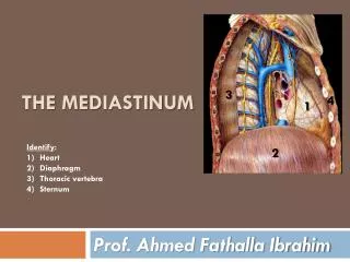
Middle mediastinum
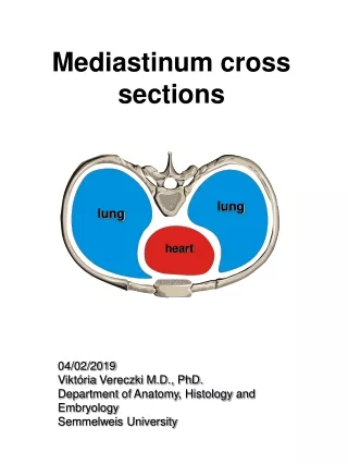
Middle mediastinum. Heart and Middle Mediastinum. superior mediastinum: [Green] Inferior Mediastinum: Below the plane passing from Sternal Angle/Angle Luise Inferior mediastinum has 3 parts: Purple: anterior mediastinum; Yellow: middle mediastinum; Blue: posterior mediastinum .


Middle mediastinum
E N D
Presentation Transcript
Heart and Middle Mediastinum • superior mediastinum: [Green] • Inferior Mediastinum: Below the plane passing from Sternal Angle/Angle Luise • Inferior mediastinum has 3 parts: • Purple: anterior mediastinum; • Yellow: middle mediastinum; • Blue: posterior mediastinum
Middle mediastinum • The middle mediastinum is centrally located in the thoracic cavity. • It contains the pericardium, heart, origins of the great vessels, various nerves, and smaller vessels.
Pericardium • The pericardium is a fibroserous sac surrounding the heart and the roots of the great vessels. • It consists of two components, the fibrous pericardium and the serous pericardium.
Fibrous pericardium • The fibrous pericardium is a cone-shaped bag with its base on the diaphragm and its apex continuous with the adventitia of the great vessels . • The base is attached to the central tendon of the diaphragm and to a small muscular area of the diaphragm on the left side. • Anteriorly, it is attached to the posterior surface of the sternum by sternopericardial ligaments.
Phrenic nerves and pericardiacophrenic vessels. • The phrenic nerves, which innervate the diaphragm and originate from spinal cord levels C3 to C5, pass through the fibrous pericardium and innervate the fibrous pericardium as they travel from their point of origin to their final destination. • Pericardiacophrenic vesselsare also located within and supply the fibrous pericardium as they pass through the thoracic cavity
Serous pericardium • Serous itself has two layers: outer – Parietal and inner – Visceral. • Pericardial space [between two layers of serous pericardium] contains a small amount of fluid that allows the heart to function in a frictionless environment
Reflections of serous pericardium • These reflections of serous pericardium occur in two locations: • 1.one superiorly, surrounding the arteries, the aorta and pulmonary trunk • 2. second more posteriorly, surrounding the veins, the superior and inferior vena cava and the pulmonary veins.
When the pericardium is opened anteriorly during surgery, a finger placed in the transverse sinus separates arteries from veins. • A hand placed under the apex of the heart and moved superiorly slips into the oblique sinus.
Transverse Sinus • Lies between • Great vessels anteriorly [aorta and pulmonary trunk] • SVC posteriorly During Open heart surgery, clamp is passed here to block the blood flow while patient is put on artificial heart-lung machine.
Oblique pericardial sinus • The zone of reflection surrounding the veins is 'J-shaped' and the cul-de-sac formed within the 'J', posterior to the left atrium, is the oblique pericardial sinus.
Pericardial effusion • Normally, only a tiny amount of fluid is present between the visceral and parietal layers of the serous pericardium. In certain situations, this space can be filled with excess fluid (pericardial effusion). • Because the fibrous pericardium is a 'relatively fixed' structure that cannot expand easily, a rapid accumulation of excess fluid within the pericardial sac compresses the heart (cardiac tamponade), resulting in biventricular failure. • Removing the fluid with a needle inserted into the pericardial sac can relieve the symptoms.
Cardiac Tamponade • Cardiac tamponade (heart compression), is a potentially lethal condition because heart volume is increasingly compromised by the fluid outside the heart but inside the pericardial cavity. • Blood in the pericardial cavity, hemopericardium, likewise produces cardiac tamponade.
Pericardiocentesis • Drainage of fluid from the pericardial cavity, pericardiocentesis, is usually necessary to relieve cardiac tamponade. • To remove the excess fluid, a wide-bore needle may be inserted through the left 5th or 6th intercostal space near the sternum.
Heart • The general shape and orientation of the heart are that of a pyramid that has fallen over and is resting on one of its sides. • Placed in the thoracic cavity, the apex of this pyramid projects forward, downward, and to the left, whereas the base is opposite the apex and faces in a posterior direction.
The sides of the pyramid consist of: • 1. Diaphragmatic (inferior) surface on which the pyramid rests • 2. Anterior (sternocostal) surface oriented anteriorly • 3.Right pulmonary surface • 4.Left pulmonary surface.
Base (posterior surface) and apex • The base of the heart is quadrilateral and directed posteriorly. It consists of: • 1. Left atrium • 2. Small portion of the right atrium • 3.Proximal parts of the great veins (superior and inferior venaecavae and the pulmonary veins)
Apex of the heart • The apex of the heart is formed by the inferolateral part of the left ventricle and is positioned deep to the left fifth intercostal space, 8-9 cm from the midsternal line.
External sulci • Internal partitions divide the heart into four chambers (i.e. two atria and two ventricles) and produce surface or external grooves referred to as sulci. • The coronary sulcuscircles the heart, separating the atria from the ventricles . • As it circles the heart, it contains the right coronary artery, the small cardiac vein, the coronary sinus, and the circumflex branch of the left coronary artery.
The anterior and posterior interventricularsulci • Separate the two ventricles- • Anterior interventricularsulcusis on the anterior surface of the heart and contains the anterior interventricular artery and the great cardiac vein • Posterior interventricularsulcusis on the diaphragmatic surface of the heart and contains the posterior interventricular artery and the middle cardiac vein.
The Heart Wall is Made Up of Three Layers From outer to inner layer • Epicardium: visceral pericardium • Myocardium: heart muscle • Endocardium: endothelium lining the heart chambers and covering the valves.
Chambers of Heart • Right atrium • Right ventricle • Left atrium • Left ventricle
Right atrium • Blood returning to the right atrium enters through one of three vessels. • These are: • 1. Superior and inferior venaecavae, which together deliver blood to the heart from the body • 2.Coronary sinus, which returns blood from the walls of the heart itself.
Right Atrium • The ear-like Right Auricle is a small, conical muscular pouch that projects from the right atrium. [it is primordial atrium represented in the adult]. • The interior of the right atrium is divided into two continuous spaces • 1.Externally, this separation is indicated by a shallow, vertical groove (the sulcusterminaliscordis) • 2. Internally, this division is indicated by the cristaterminalis • Its walls are covered by ridges called the musculipectinati (pectinate muscles), which fan out from the crista like the 'teeth of a comb'.
Right ventricle • In the anatomic position, the right ventricle forms most of the anterior surface of the heart and a portion of the diaphragmatic surface. • Blood entering the right ventricle from the right atrium therefore moves in a horizontal and forward direction. • The outflow tract of the right ventricle, which leads to the pulmonary trunk, is the conusarteriosus (infundibulum).
Right Ventricle • The interior of the right ventricle has irregular muscular elevations called TrabeculaeCarneae. • The inflow part of the right ventricle receives blood from the right atrium through the Tricuspid orifice, located posterior to the body of the sternum at the level of the 4th and 5th intercostal spaces.
Papillary muscles • The Papillary muscles form conical projections with their bases attached to the ventricular wall and tendinous cords arising from their apices. There are usually three papillary muscles (anterior, posterior, and septal) in the right ventricle that correspond in name to the cusps of the tricuspid valve. • The SeptomarginalTrabecula (moderator band) is a curved muscular bundle that runs from the inferior part of the interventricular septum to the base of the anterior papillary muscle
Tricuspid valve • The right atrioventricular orifice is closed during ventricular contraction by the tricuspid valve (right atrioventricular valve), which is so-named because it usually consists of three cusps or leaflets . • The base of each cusp is secured to the fibrous ring that surrounds the atrioventricular orifice. This fibrous ring helps to maintain the shape of the opening. • The naming of the three cusps, the anterior, septal, and posterior cusps, is based on their relative position in the right ventricle
Pulmonary Valve • The Pulmonary Valve at the apex of the conusartereosus is at the level of the left 3rd costal cartilage. Each of the 3 semilunar cusps of the pulmonary valve (anterior, right, and left) is concave when viewed superiorly.
Inter Ventricular Septum • The Interventricular Septum, composed of membranous and muscular parts, is a strong, obliquely placed partition between the right and the left ventricles, forming part of the walls of each.
Left atrium • The left atrium forms most of the base or posterior surface of the heart. • A slightly thicker wall than that of the right atrium. • A larger smooth-walled part and a smaller muscular auricle containing pectinate muscles. • Four pulmonary veins (two superior and two inferior) entering its posterior wall.
Left ventricle • It contributes to the anterior, diaphragmatic, and left pulmonary surfaces of the heart, and forms the apex. • A double-leaflet Mitral Valve that guards the left AV orifice, Blood enters the ventricle through the left atrioventricular orifice and flows in a forward direction to the apex. • Walls that are covered with thick muscular ridges, trabeculaecarnae, that are finer and more numerous than those in the right ventricle. • Anterior and posterior papillary muscles that are larger than those in the right ventricle.
Aortic Valve • The Aortic Valve, obliquely placed, is located posterior to the left side of the sternum at the level of the 3rd intercostal space. • The Aortic Sinuses are the spaces at the origin of the ascending aorta between the dilated wall of the vessel and each cusp of the aortic (semilunar) valve. • The mouth of the right coronary artery is in the right aortic sinus and the mouth of the left coronary artery is in the left aortic sinus and no artery arises from the posterior [non coronary] aortic sinus
Fibrous skeleton of heart • The cardiac skeleton is a collection of dense, fibrous connective tissue in the form of four rings with interconnecting areas in a plane between the atria and the ventricles. • The four rings of the cardiac skeleton surround the two atrioventricular orifices, the aortic orifice and opening of the pulmonary trunks.
Where to listen for heart sounds • The tricuspid valve is heard just to the left of the lower part of the sternum near the fifth intercostal space. • The mitral valve is heard over the apex of the heart in the left fifth intercostalspace at the midclavicular line. • The pulmonary valve is heard over the medial end of the left second intercostalspace. • The aortic valve is heard over the medial end of the right second intercostalspace.
Coronary Circulation • Two coronary arteries arise from the aortic sinuses in the initial portion of the ascending aorta and supply the muscle and other tissues of the heart. • They circle the heart in the coronary sulcus, with marginal and interventricular branches, in the interventricularsulci, converging toward the apex of the heart