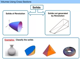
Understanding Mediastinal Divisions and Structures in Anatomy
400 likes | 737 Vues
Explore the divisions of the mediastinum such as superior, anterior, middle, and posterior, and learn about key structures including thymus, great veins, great arteries, phrenic nerve, and more. Take a detailed look through cross-sectional images and radiological scans.

Understanding Mediastinal Divisions and Structures in Anatomy
E N D
Presentation Transcript
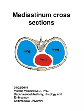
lung lung heart Mediastinum cross sections 04/02/2019 Viktória Vereczki M.D., PhD. Department of Anatomy, Histology and Embryology Semmelweis University
Divisions of mediastinum in Gray’s: Superior mediastinum Anterior mediastinum Middle mediastinum Posterior mediastinum in Réthelyi-Szentágothai : Anterior mediastinum -supracardiac -cardiac Posterior mediastinum
lung lung heart Mediastinum Mediastinum anterius -mediastinum supracardiacum: 1. thymus –corpus adiposum retrosternale 2. great veins 3. great arteries 4. n. phrenicus 5. n. vagus 6. n. laryngeus recurrens 7. trachea -mediastinum cardiacum: pericardium and heart Mediastinum posterius 1. oesophagus 2. aorta thoracica 3. aa. intercostales 4. ductus thoracicus 5. v. azygos, v. hemiazygos, v. hemiazygos accessoria 7. truncus sympathicus 8. nn. splanchnicus minor et major 9. nn. Intercostales 10. nn. vagi
Mediastinum anterius -mediastinum supracardiacum: 0. endothoracic fascia 1. thymus –corpus adiposum retrosternale 2. great veins 3. great arteries 4. n. phrenicus 5. n. vagus 6. n. laryngeus recurrens 7. trachea
Mediastinum anterius -mediastinum supracardiacum: 0. endothoracic fascia 1. thymus –corpus adiposum retrosternale 2. great veins 3. great arteries 4. n. phrenicus 5. n. vagus 6. n. laryngeus recurrens 7. trachea
Thymus The neonatal thymus: From a 9 year old female from 80 old male
Great veins Brachiocephalic veins
Great arteries a. carotis comm. sin.
Vagus nerve- recurrent laryngeal nerve Ligamnetum arteriosum
Trachea (+ nodi lymphatici paratracheales, bronchomediatinales) Th4
Laryngo-bronchoscopia Carina tracheae Plica vocalis
Mediastinum posterius –right view 1. oesophagus 2. aorta thoracica 3. aa. intercostales 4. ductus thoracicus 5. v. azygos, v. hemiazygos, v. hemiazygos accessoria 7. truncus sympathicus 8. nn. splanchnicus minor et major 9. nn. intercostales 10. nn. vagi
Mediastinum posterius –left view 1. oesophagus 2. aorta thoracica 3. aa. intercostales 4. ductus thoracicus 5. v. azygos, v. hemiazygos, v. hemiazygos accessoria 7. truncus sympathicus 8. nn. Splanchnicus minor et major 9. nn. Intercostales 10. nn. vagi
CT in the supracardial mediastinum At the 1st costochondral junction • Manubrium • Right brachiocephalictrunc • Right brachiocephalicvein • Trachea • Scapula • 6. leftbrachiocphalicvein • 7. Leftcommoncarotidartery • 8. Leftsubclavianartery • 9. esophagus
CT in the supracardial mediastinum T3 • brachiocephalic vein • Pretracheal space • Trachea • esophagus • Pectoralis major • Left subclavian artery Left subclavian artery • scapula
CT in the supracardial mediastinum T4 • 2. Superiorvenacava • 3. Pretrachealspace • 4. Trachea • 5. Arch of aorta • 6. esophagus
CT in the supracardial mediastinum T6 • 1. Ascending aorta • 2. Superiorvenacava • 3. Right pulmonaryartery • 4. Right principalbronhc • 5. Subcarinalspace • 6. Esophagus • 7. Azygosvein • 8.Trunk of pulmonaryartery • 9. Leftsuperiorpulmonaryvein • 10. Leftprincipalbronchus • 11. Inferiorbranch of theleftpulmonaryartery • 12. Descending aorta
CT in the supracardial mediastinum T7 • 1. Aorticroot • 2. Leftatrium • 3. Esophagus • 4. Azygosvein • 5. Right ventricular outflow track • 6. Leftinferiorpulmonaryvein • 7. Descending aorta
Thankyou! References: Gray’s Anatomy, 41st ed. 2016 M. Réthelyi and J. Szentágothai: Functional Anatomy 2018