Myopathy
Explore the differences between lower and upper motor neuron weakness, learn about symptoms of muscle disease, understand diagnostic procedures like ENG/EMG and muscle biopsy, and discover the management strategies for muscular dystrophies like Duchenne and Becker.

Myopathy
E N D
Presentation Transcript
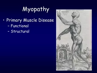
Myopathy • Primary Muscle Disease • Functional • Structural
Lower vs. UpperMotor Neuron Weakness *Disuse atrophy can develop after initial presentation
Distinguishing Lower Motor Weakness from Muscle Weakness • Weakness due to neuropathy: lower motor neuron disease. • Weakness due to myopathy: nerve function intact.
Symptoms of Muscle Disease • Negative • Weakness • Proximal and symmetric • Waddling gait; difficulty of rising from sitting, climbing stairs; getting up from a squatted position, Gower’s sign • Fatigue; exercise intolerance • Metabolic, mitochondrial • Atrophy • Positive • Myalgia • Cramp • Contractures • Increased lordosis, scoliosis • Myotonia • Impaired relaxation of muscle after forceful voluntary contraction • Due to repetitive depolarization of the muscle membrane • Myoglobinuria • Dark colored urine • Excessive release of myoglobin from muscle • Metabolic-toxic-idiopathic • Pseudohypertrophy • Tendon reflexes are normal or depressed from A Kornberg MD
Key questions • Constant or episodic symptoms • Age at onset • Is there a family history • Acute,subacute, chronic • Life long • Congenital • Progressive or non-progressive • Proximal-distal symptoms • Cranial involvement • Triggering factors • Exercise • Carbonhydrate • Relieved by exercise • Systemic signs • Rash, dark/red urine, cardiac, cataract, neuropathy, mental retardation
Differential Diagnosis Lab • CK • Electrolytes • Serum myoglobin • Serum kreatinin, BUN • Complete Blood Count • ESR • Thyroid function tests • AST • Urinalysis • Myoglobinuria
Diagnosis of muscle diseases • Creatinine kinase levels increased in many myopathies • sign of muscle fiber necrosis • ENG / EMG: differentiation between neurogenic and myogenic weakness • Muscle biopsy: signs of muscle fiber abnormality, inflammation, immunostaining of muscle constituents • Genetic testing • EKG-Echocardiography
Classification of Myopathies • Hereditary muscle diseases • Muscle dystrophies • Myotonias and Channelopathies • Congenital Myopathies • Metabolic myopathies • Mitochondrial myopathies • Acquired muscle diseases • Inflammatory myopathies • Idiopathic, infectious • Endocrine myopathies • Myopathies associated with other systemic illness • Drug induced/toxic myopathies
Muscular Dystrophies • Hereditary • Progressive • Pseudohypertrophy +++ • Muscles replaced by fat • Very High CK • Genetic defect of proteins constituting the sarcolemma-associated cytoskeleton system • Dystrophinopathies and others
Muscular Dystrophies • Dystrophinopaties • Duchenne, Becker • Limb Girdle dystrophies • Fascioscapulohumeral dystrophy • Oculopharyngeal dystrophy • Distal muscular dystrophies • Myotonic dystrophies
Muscular Dystrophies-Duchenne • X linked recessive • Absence of dystrophinprotein • Slow to reach motor milestones • Symptoms by age 5 • All walk, may never run • End up in wheelchair by age 12 • Frequently mildly mentally retarded • Life expectancy < 20 years with death related to respiratory failure or cardiomyopathy • Dx- Genetic, muscle biopsy
Duchenne muscular dystrophy Normal Duchenne dystrophy • Diagnosis: • Lack of immunostaining of dystrophin in muscle biopsy specimen • Demonstration of deletion in the dystrophin gene
Muscular Dystrophies-Becker • Less common than Duchenne • X-linked recessive • Later Onset • 10 years • Most patients ambulant until 3rd-4th decade • Death from heart failure due to dilated cardiomyopathy-late 40’s • Mental retardation is rare
Management of DMD/BMD • Annual echocardiography • Cardiomyopathy • ACE inhibitor and/or beta blocker....digoxin • Cardiomyopathy and decreased pulmonary function + • Pneumoccal vaccine • Annual influenza vaccine • Anesthesia complications...... • Consider TIVA • Susceptibility to malign hyperthermia • Rhabdomyolisis, hyperkalemia, cardiac arrest • Succinylcholine, halothane, isoflurane, sevoflurane • Reaction to inhaled anesthetics, muscle relaxants, hypoventilation, atelectasis, cardiac arrhytmias, heart failure, difficulty weaning from mechanical ventilation,
Management of DMD/BMD • Low dose daily prednol - deflazacort in DMD • İncrease of muscle strength-maximum at 3 months • Prolongs independent ambulation • İmprovement of FVC • Preserves cardiac function • Increases osteoporosis • AAN recommendations • Prednol 0,75 mg/kg/day for patients over the age of 5 years • Watch for weight gain, cushingoid appearance, fractures, gastrointestinal symptoms..... • Decrease to 0,5 then 0,3 mg/kg/day if weight gain occurs • Deflazacort 0,9 mg/kg/day • Watch for weight gain and cataracts
Management of DMD/BMD • Vitamin D supplement • If serum vitamin D level less than 20ng/ml • Nutritionist • Avoid obesity • Physical Therapy, orthopedic devices • Gene Therapy • Aminoglycosides, PTC124, Creatine
Facioscapulohumeral dystrophy • Prevalence: 1 in 20,000 • Most common after DMD and BMD • Autosomal dominant • Age of onset: before 20 • Sensorineural hearing loss
Facioscapulohumeral dystrophy • myopathic face, • İnability to smile, whistle or close eyes • Life span is not significantly affected Charcot
Facioscapulohumeral dystrophy • Progressive muscular weakness and atrophy involving the face, scapular, proximal arm and peroneal muscles winging of the scapula
Oculopharyngeal Muscular Dystrophy • 5’th-6’th decade • Extraocular muscle weakness • Ptosis • Dysphagia • Later facial and proximal muscle weakness • Autosomal Dominant
Emery Dreifuss Muscular DystrophyEDMD • Childhood- adolescence • Slowly progressive • Contractures before weakness • Neck extansors, elbows, ankles • Cardiomyopathy • Atrial arrhythmias and syncope From: A Kornberg Emery-Dreifuss: Rigid spine
Limb-girdle dystrophies • Autosomal Dominant (10%) • LGMD-1 • Autosomal Recessive (90%) • LGMD-2 • Symptoms between 2-40 • High CK • Slow progressive proximal muscle weakness • Onset variable • Spares the face
Limb-girdle dystrophies • Causes: • Sarcoglycanopathies • α, β, γ, δ sarcoglycans • Clinically often indistinguishable from Duchenne-dystrophy • scapular winging more pronounced, hamstrings and quadriceps equally affected • Calpain - 3 mutation • Dysferlin deficiency etc. • Caveolin deficiency (AD)
Sarcoglycanopathies Sarcoglycanopathy Normal
Myotonic dystrophy • Prevalence: 1 in 8000 • Cause: CTG repeat expansion in a gene on chr. 19 • Autosomal dominant inheritance, with anticipation • Multisystemic disease: • Myotonia: hyperexcitability of muscle membrane inability of quick muscle relaxation • Progressive muscular weakness and wasting, most prominent in cranial and distal muscles • Cataracts, frontal balding, testicular atrophy • Cardiac abnormalities, mental retardation, endocrinopathy
Periodic paralysis • Characterized by episodes of flaccid muscle weakness occurring at irregular intervals • Strength is normal between attacks • General characteristics : • Hereditary; • Most are associated with alteration in serum potassium levels; • Hypokalemic, hyperkalemic, normokalemic • Myotonia sometimes coexists; • Both myotonia and PP result from defective ion channels.
Periodic paralysis • The physiologic basis of flaccid weakness is inexcitability of the muscle membrane • (ie, sarcolemma).
Myotonia congenita • Mutation in the muscle Cl gene • Autosomal dominant • Thomsen • Autosomal recessive • Becker • Symptoms: • Myotonia (hyperexcitability of the muscle membrane): muscle stiffness and abnormal muscle relaxation, warm-up phenomenon • Hypertrophied muscles • Therapy: phenytoin, mexiletin
Thyrotoxic periodic paralyses • Common in adults aged 20-40 years. • Hyperinsulinemia, a carbohydrate load, and exercise are important in precipitating paralytic attacks. • Attacks last hours to days.
Congenital Myopathies • Age of onset in childhood • Occasionally adults • Familial • Occasionally sporadic • Floppy infant • Hypotonia, muscle weakness • Structered • Central core disease • Multi-minicore disease • Myotubular and centronuclear myopathies • X-linked myotubular myopathy • Centronuclear myopathy • Nemaline myopathy • Actin aggregate myopathy • Desminopathy • Crystallinopathy • Hyaline body myopathy • Unstructured • Congenital fiber type disproportion
Metabolic Myopathies Failed Energy Production • Classification of metabolic myopathies • Carbonhydrate processing disorders • Fat-processing disorders • Mitochondrial disorders
Metabolic Myopathies • Dynamic rather then static symptoms • Cramps, pain, exercise intolerance rather than fixed symptoms • A history of weakness, pain and/or myoglobinuria provoked by short duration high intensity exercise, • Lifting heavy weight, swimming, climbing stairs, running • Exercise intolerance and myoglobinuria • Glycolytic pathway defect • Episodic weakness in association with fever, after prolonged mild exercise or prolonged fasting • After 1 hour • Walking • Lipid metabolism defects
Metabolic Myopathies Laboratory • Urinary • Ketons • Myoglobin • Dicarboxylic acids • Acylglycins • Serum • Lactate • Pyruvate • Lactic acid dehydrogenase • Uric acid • Free and total carnitine • Ketones • Glucose • Ammonia • Myoglobin • Liver transaminases • Potassium, calcium, phosphate • Creatinine • Acylcarnitine CK at rest and during an episode
Mitochondrial Myopathies • Mitochondria • Main source of metabolic energy
Mitochondrial Myopathies • Nuclear DNA defects • Leigh, MNGIE, MDS • Mitochondrial DNA defects
Idiopathic Inflammatory Myopathies • Clinical Classification • Polymyositis (PM) • Dermatomyositis (DM) • Inclusion Body Myositis (IBM) • Juvenile Dermatomyositis • Myositis associated with malignancy • Myositis associated with collagen vascular disease Bohan & Peter (1975). NEJM. Tanimoto et al. (1995). J Rheumatology.
Polymyositis / Dermatomyositis • Diagnostic criteria • Proximal muscle weakness • Elevated serum CK • Myopathic changes on EMG • Muscle biopsy demonstrating lymphocytic inflammation • Dermatomyositis: Skin rash as well as criteria above • Definitive diagnosis with four criteria having been met Bohan & Peter (1975). NEJM.
Polymyositis-Dermatomyositis • Painful muscles (60%) • Polymyositis: • Cell-mediated immune response against muscle fibers • Less often paraneoplastic than DM • Cardiac abnormalities • Dermatomyositis: • Humorally mediated autoimmune microangiopathy affecting the muscles and skin • Rash on the face, neck • Periorbital oedema • Dysphagia • Cardiac abnormalities, interstitial lung disease • May be paraneoplastic
Treatment for DM, PM • Immunosuppression • long-term treatment with corticosteroids (1 mg/day) • Mtx-Imuran...
Inclusion Body Myositis • Mainly affects older males • Symptoms begin insidiously and progress slowly • Symptoms are often present 5-6 years before diagnosis • Finger flexors-quadriceps • May include focal, distal or asymmetric weakness • Neurogenic or mixed neurogenic / myopathic changes on EMG • Dysphagia is noted in more than 20% of patient Wortmann RL. Primer on Rheum Dis. 12th edition. 2001:369–376.
Dermatomyositis B cells, macrophages CD4 T cells Decreased capillaries Perifascicular atrophy Perivascular infiltrate Polymyositis Mononuclear cells CD8 T cells Endomysial infiltrate Myonecrosis Patchy, focal Muscle Biopsy:Histology and Immunochemistry • IBM • Same as PM; also: • Rimmed vacuoles • Eosinophilic cytoplasmic inclusions Rolak LA. Neurology Secrets. 2005: 63-7.
Infectious causes of myopathy • Trichinosis • Cysticercosis • Toxoplasmosis • HIV • Coxsackie A and B • Influenza • Lyme • Staphylococcus aeurus
Endocrine myopathies • Endocrine myopathies • Thyrotoxic myopathy • Hypothyroidism • Hyperparathyroidism • Adrenal insufficiency • Hypokalemia