Download
1 / 68
1.05k likes | 3.74k Vues
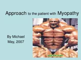
Approach to the patient with Myopathy. By Michael May, 2007. Outline. Basics Clinical manifestations Diagnostic approach Individual myopathies Approach to the patient. Skeletal muscle. Composed of muscle fibers that contract when stimulated by a motor neuron

E N D
Approach to the patient with Myopathy By Michael May, 2007
Outline • Basics • Clinical manifestations • Diagnostic approach • Individual myopathies • Approach to the patient
Skeletal muscle • Composed of muscle fibers that contract when stimulated by a motor neuron • Skeletal muscle: covered by connective tissue sheath( epimysium) : composed of many columns( fascicles) • Fascicles: surrounded by perimysium :composed of many muscle fibers • Muscle fibers: covered by cell membrane ( sarcolemma) and surrounded by endomysium. :anatomic and physiologic unit . Two types : 1. Type - I / slow twitch/ red fibers : richer in oxidative but poorer in glycolytic enzymes. 2. Type- II/ fast twitch/ white fibers :richer in glycolytic but poorer in oxidative enzymes. : composed of nuclei, cytoplasm ( sarcoplasm), myofibrils, ribosomes, mitochondria, stored fat, many enzymes, glycogen, myoglobin.
Cont,d • Myofibril : longitudinally oriented interdigitating filaments of contractile proteins ( Actin, Myosin) & regulatory proteins ( Troponin, tropomyosine, nebuline). : Thick filaments: composed of protein, Myosin. : Thin filaments: composed of protein , Actin. : enveloped by a membranous net ( sarcoplasmic reticulum) • Motor unit : each somatic motor neuron together with all of the muscle fibers it innervates. : each neuron innervates muscle fibers of one type only. : Neuromuscular junction( NMJ) : junction of the terminal of the motor neuron with the muscle fiber. : Motor end plate: specialized region of the sarcolemma of the muscle fiber at the NMJ.
Cont,dMotor unit courtesy : Prof. Janzer
Cont,d • Muscle contraction: sliding of thin filaments over and b/n thick filaments by the action of numerous cross bridges that extend out from the myosine toward the actin. • : muscle fiber stimulation by motor neuron → ca+2 moves from its store site in the sarcoplasmic reticulum to the sarcoplasm→ attaches to troponine → conformational change that moves the troponin complex and its attached tropomyosine out of the way so that cross bridges can attach to actin→ sliding of filaments and thus muscle tension and shortening. ▪ Energy source : fatty acids , glycogen and blood glucose, aminoacids : skeletal muscles generate ATP through anaerobic and aerobic respirations and phosphate groups donated by creatine phosphate. ATP : serves as the immediate source of energy for 1. movement of cross bridges for muscle contraction 2. pumping of ca+2 in to the sarcoplasmic reticulum for muscle relaxation.
Cont,d • Mitochondria : plays key role in energy production • Anaerobic respiration : major source of energy during exercise Glucose → Lactic acid + ATP • Aerobic respiration : lipids – important sources of energy during rest & during prolonged submaximal exercise VLDL Triglycerides → fatty acids → activated fatty acids/ acyl-coA/ ↓ CPT-I linked with carnitine ↓ mitochondria ↓ CPT-II ATP ← Beta oxidation ← acyl-coA + carnitine
Myopathy • Defn. : disorders with structural changes or functional impairment of a muscle; unrelated to any disorder of innervation or NMJ. • Classification : 1. Acquired 2. Hereditary/ genetic : mutations or deletions of genes coding for parts of a muscle ( filament proteins, mitochondrial enzymes, sarcoplasmic reticulum, specialized channels for entry of ca+2 , Na+2, Cl- , K+1 transverse tubules, sarcolemma ) : affect structure or biochemical processes which convert chemical energy derived from cell metabolism in to mechanical energy in a controlled manner.
Causes • Inflammatory • Metabolic • Toxic • Congenital /inherited • Endocrinopathies • Chanellopathies • Muscular dystrophies
Clinical presentation • Muscle weakness • Fatigue • Muscle pain( myalgia), cramps and stiffness • Muscle contracture • myoglobinuria • Myotonia • Muscle enlargement and atrophy • Manifestations of specific illnesses
Muscle weakness • Symptoms: Proximal – difficulty in combing hair, getting up from a chair, climbing stairs, getting up from a squatting position. :Distal – difficulty buttoning, writing, knitting. • Types: Intermittent ( periodic)- proximal > distal ( limb- girdle pattern ) - HypoKPP, HyperKPP, paramyotonia congenita - metabolic energy deficiencies of glycolysis - abnormalities in fatty acid metabolism - myasthenia gravis: NCS~ repetitive nerve stimulation decremental response : Persistent ( fixed) - most muscle disorders - limb - girdle pattern /face spared/ is common - some patterns are restricted to certain diseases.
Inability to maintain or sustain a force fatigue • Pathologic fatiguability -Disorders of neuromuscular transmission, disorders altering energy metabolism ( glycolysis, lipid metabolism), disorders in mitochondrial energy production - Chronic myopathies - Accompanied by abnormal clinical or laboratory findings • Asthenia - a type of fatigue caused by excess tirdeness or lack of energy - a tendency to avoid physical activities - complaints of daytime sleepiness - necessity for frequent naps - difficulty concentrating on activities such as reading - feelings of overwhelming stress & depression
Muscle pain (myalgia), cramps, and stiffness- not a feature of most primary muscle diseases • Cramp: painful, involuntary, localized, muscle contraction with a visible or palpable hardening of the muscle. : often occur in neurogenic disorders/ motor neuron diseases, radiculopathies, polyneuropathies/ : common in patients with Duchenne MD : firing of MUAPs at a rate of 40-60 Hz, with abrupt onset & cessation • Myalgia: localized or generalized and may be accompanied by tenderness & swelling. :should be differentiated from myofascial pain syndromes • Fibromyalgia▪polymyalgia rheumatica - have specific trigger points - in patients > 50yrs of age - easy fatiguability - stiffness & pain in the shoulders, - sleep disturbances lower back, hips, and thighs - serum CK & ESR are normal - ESR elevated - CK, EMG & muscle biopsy are N
Cont,d stiffness • Uncommon • Joint inflammation • Hyperexcitable motor nerves( spinal cord, peripheral nerves) contracture • Muscle unable to relax after after an active muscle contraction • EMG shows complete electrical silence ( firing of motor neurons in a cramp) • Uncommon in most muscle diseases • Fixed contracture occurs early in patients with Emery- Dreifuss MD & Bethlem myopathy.
Cont,d myoglobinuria ▪ dark or dark- brown urine Myotonia • prolonged muscle contraction followed by slow muscle relaxation • voluntary contraction, mechanical stimulation ( percussion myotonia) • DM, myotonia congenita, paramyotonia congenita Muscle enlargement and atrophy • Size of muscle is usually not affected. • Enlarged cuff muscles are typical Duchenne & Becker MDs. • Can also result from infiltration by sarcoid granulomas , amyloid deposits, bacterial and parasitic infections, and focal myositis.
Laboratory evaluation • Diagnose myopathy • Serum enzymes - ALT, AST, LDH, Aldolase : found in both skeletal muscle & liver : elevated GGT helps to establish a liver origin. - CK ( CKMM) :preferred enzyme to measure in the evaluation of myopathies. • Electrodiagnostic studies - NCS, Repetitive nerve stimulation : help to differentiate myopathies from neuropathies & NMJ disorders. - EMG : diagnose myopathy, helps also to choose an appropriately affected muscle for biopsy. - Myopathic EMG: low amplitude, short duration & polyphasic MUPs - inflammatory myopathies : increased spontaneous activity, irritability on needle placement. - myotonic discharges : sustained run of positive sharp waves : sustained run of negative spikes
Lab. Cont,d • Diagnose specific types of myopathies • Forearm exercise test - place a small indwelling catheter in to an antecubital vein & obtain baseline blood sample for lactic acid and ammonia. - the fore arm muscles are exercised by asking the patient to vigorously squeeze a sphygmomanometer bulb for 1 min. -blood is then obtained at intervals of 1,2,4,6 & 10 min. for comparison with the baseline. Both rise with exercise. - Glycolytic defects : lactic acid rise is absent or below normal while rise in ammonia will reach control values. - myoadenylate deaminase deficiency: selective failure to increase ammonia.
Lab. Cont,d • DNA analysis - for definite diagnosis of certain muscle disorders associated with gene defects ( deletions or mutations ). • Muscle biopsy - safe diagnostic procedure in establishing the final diagnosis of suspected myopathy. - different techniques of microscopic evaluation ( histochemistry, immunohistochemistry with a battery of antibodies, electron microscopy) - site :muscle selected for biopsy should have mild to moderate muscle weakness. : not performed on a muscle that has been injured by a previous trauma, injections, EMG needles( within 4-6 weeks after EMG ) - common muscles used for biopsy : proximal – Biceps, Triceps, Quadriceps : Distal – Extensor carpi radialis, Anterior tibialis
Approach to the patient • Emergency: manage acute life threatening complications Respiratory insufficiency : mechanical ventilator Dysphagia : endotracheal intubation Rhabdomyolysis : hydration, diuresis Heart block : pace maker insertion HypoKPP : iv. Or oral potassium replacement • Non-emergency : identify a disorder as a myopathy : identify a specific etiology for the myopathy • History - symptoms : muscle weakness, presence or absence of sensory complaints , acuity of symptom onset - family history of muscle weakness, frontal baldness - personal history of autoimmune disease , endocrinopathy, renal insufficiency, alcoholism - Previous history of severe weakness, particularly any that occurred after exercise, exposure to cold temperatures, eating high CHO diets
Approach cont,d - history of medication use • Physical examination - objective weakness - fever ( polymyositis ) - muscle tenderness - muscle mass : atrophy is a very late sign - skin examination : Gottron’s papule, heliotrope rash - level of consciousness : usually normal - sensory perception : normal - DTR : usually normal : may be absent or diminished in HypoKPP • Laboratory tests : CBC, ESR, CK isoenzymes, electrolytes, U/A serum myoglobin, RFT, TFT Definitive diagnosis : EMG, Genetic testing, ANA, Muscle enzymes, muscle biopsy
Muscular dystrophies • Group of inherited disorders characterized by progressive degeneration of groups of muscles, sometimes with involvement of the heart muscle or conducting tissue, and other parts of the nervous system. • Classified based on the age at onset, distribution of affected muscles and pattern of inheritance. Duchenne muscular dystrophy • Inheritance- X- linked recessive disorder • Defective gene- Dystrophin • Onset- usually b/n 3-5yrs age • C/F – progressive weakness of the girdle muscles - difficulty running , jumping, hopping, unable to get up from the floor (Gower’s maneuver) - toe walking is associated with lordotic posture - contractures( hip, knee, elbow, wrist) with chest deformities →severe pulmonary infections → death at age 16-18yrs
Cont,d Others : cardiomyopathy , mental retardation • Lab. – Serum CK : elevated 20-100x normal - EMG : myopathic features - Muscle biopsy: muscle fibers of varying size as well as small groups of necrotic and regenerating fibers. : deficiency of dystrophin seen on western blot analysis & immunohistochemical staining. - DNA analysis : mutation of gene that encodes dystrophin • Treatment : prednisolone 0.75mg/kg/day increases muscle strength & slows the progression of disease for up to 3 yrs.
Becker muscular dystrophy • Inheritance – X- linked recessive disorder • Defective gene – dystrophin • Onset- experience difficulty b/n 5- 15yrs of age • C/F – proximal muscles especially of lower extremities are prominently involved. - hypertrophy of muscles , particularly the calves, is an early & prominent finding. - cardiomyopathy may occur , MR is less common • Lab. – CK : elevated - EMG : myopathic - muscle biopsy : similar to DMD : reduced amount or abnormality of dystrophin( Dx) - DNA analysis : deletions or duplications( Dx) • Treatment – supportive • Survival : survive in to the 4th to 5th decade
Limb- girdle muscular dystrophy/ LGMD / • Inheritance : Autosomal dominant/ recessive • Defective gene : several genes • M:F – 1:1 • Onset – late 1st to 4th decade • C/F – progressive weakness of pelvic & shoulder girdle muscles - diaphragmatic weakness & cardiomyopathy may also occur - intellectual function is intact • Treatment - supportive
Emery- Dreifuss muscular dystrophy/EDMD/ • Inheritance- X-linked recessive/ Autosomal dominant • Defective gene : Emerin/ Lamins A/C • Onset – early childhood & teenage years • C/F – prominent and early contractures ( elbows, neck) often preceding muscle weakness. - muscle weakness is in a limb- girdle distribution - dilated cardiomyopathy may occur and may result in sudden death, arrhythmia, & conduction defects. ▪ Lab. - CK : 2-10x ed - EMG : Myopathic - Biopsy : non-specific dystrophic features • Treatment - supportive : Ambulatory aid : manage cardiomyopathy & arrhythmia
Fascioscapulohumeral /FSH/ muscular dystrophy • Inheritance: AD • Onset : childhood or young adulthood • Defective gene: deletion, distal 4q • C/F- facial weakness: initial manifestation - weakness of shoulder girdle muscles : weak arm elevation : scapular winging - weak wrist extension > wrist flexion - foot drop : weakness of anterior compartment muscles of the legs - weakness of the pelvic girdle muscles : 20% - other organ ( rarely) : labile HTN, nerve deafness • Lab. – CK : N or elevated - EMG: myopathic pattern - biopsy: non-specific features of myopathy • Treatment – no specific treatment is available - ankle foot orthoses may help for foot drops - scapular stabilization procedures may improve scapular winging
Oculopharyngeal dystrophy • Inheritance: AD with complete penetrance • Defective gene: expansion, poly-A-RNA binding protein • Onset – usually late onset ( 4th – 5th decade ) • C/F – progressive external ophthalmoplegia ( slowly progressive ptosis, limitation of eye movements with sparing of pupillary rxns. - dysphagia : can be life threatening : may result in repeated episodes of aspiration - mild weakness of the neck and extremities • Lab. – EMG: myopathic features - CK : 2-3x N - biopsy : distinct features – presence of tubular filaments in muscle cell nuclei. • Treatment- Dysphagia : cricopharyngeal myotomy may improve swallowing - Ptosis : eyelid crutches may improve vision
Distal myopathies • Notable for their preferential distal distribution of muscle weakness in contrast to most muscle conditions associated with proximal weakness • Four types : mode of inheritance, age of onset, pattern of weakness 1. Welander DM : AD 2. Tibial MD : AD - late onset, usually after age 40; start in the hands 3. Nonanka DM: AR 4. Miyoshi myopathy : AR - early onset in late teens or twenties; start in the lower limbs • Lab. – CK : only slightly elevated except in Miyoshi myopathy - Biopsy : non- specific dystrophic changes - EMG : myopathic • Treatment – occupational therapy for loss of hand function - Ankle - foot orthoses to support distal lower limbs
Myotonic dystrophy/ DM / • Inheritance : AD • Defective gene: two types with distinct molecular genetic defects -DM1 : expansion CTG repeat - DM2 ( proximal myotonic myopathy – PROMM ): CCTG repeat • C/F – myotonia : usually appears by age 5 yrs - Hatchet- faced appearance: temporalis , masseter , facial muscle atrophy & weakness - frontal baldness in men - foot drop : ankle dorsiflexor weakness - weakness of wrist extensors , finger extensors, & intrinsic hand muscles - early involvement of neck muscle flexors, sternocleidomastoids - dysarthritic speech, nasal voice, swallowing problems due to palatal , pharyngeal, and tongue involvement - respiratory insufficiency : diaphragm & intercostal muscle involvement - cardiac disturbances : conduction block with sudden death : CHF from cor pulmonale 2ry to respiratory failure
Cont,d Hatchet-faced appearance
Cont,d • - other system manifestations : intellectual impairment, hypersomnia, cataract, gonadal atrophy, insulin resistance, reduced esophageal & colonic motility • Lab. – Dx ; usually based on clinical findings - CK : N or mildly elevated - EMG : evidence of myotonia - Biopsy : atrophy which selectively involves type – 1 fibers in 50% • Treatment – treat myotonia : membrane stabilizing agents : phenytoin is preferred - pacemaker for advanced conduction block - molded ankle foot - orthoses help prevent foot drop in patients with distal lower extremity weakness.
Congenital myopathy • Rare disorders distinguished from muscular dystrophies by the presence of specific histochemical & structural abnormalities in muscle fibers. • Onset : infancy or childhood • Three types: pattern of inheritance & type of structural abnormality in muscle fibers -central core disease : AD - Nemaline (rod ) myopathy: AD - Myotubular( centronuclear ) myopathy : AD , XR • C/F - progressive muscle weakness ( proximal> distal, legs> arms) & limpness, hypotonia & delayed milestones /walking/ - skeletal deformities (kyphoscoliosis, club foot, hip dislocation) • Lab. - CK: usually N or slightly elevated - EMG : myopathic/ mostly/; positive sharp waves, myotonic discharges - Biopsy : features specific to each type • Treatment – no specific treatment
Disorders of muscle membrane excitability/chanellopathies/ • Inherited abnormalities (mutations) of the Na+, Ca+2, K+, Cl- ion channels in striated skeletal muscles. • C/F- various syndromes of familial periodic paralysis affecting proximal muscles more than distal, mostly sparing ocular , bulbar and respiratory muscles. : long term repeated attacks may result in fixed proximal weakness. - myotonia : paradoxic in PC (aggravated by exercise ) :HyperKPP ( K+ -aggravated myotonia) , few cases of HypoKPP • Inheritance – AD except few sporadic cases • Recognized by their : clinical characteristics : provocation by exercise, eating , cold exposure, and associated changes : serum potassium concentration during an attack
HypoKPP Attacks may stay for as long as 24hrs Precipitated by rest following exercise, meals high in CHO, Na+ Biopsy shows single or multiple centrally placed vacuoles Rx of acute paralysis - K+ supplementation (oral or IV ) Prevention of recurrent attacks -low CHO, Na+ diet -avoid intense exercise -K+ -sparing diuretics -Acetazolamide 125-1000mg/day HyperKPP Attacks are brief and mild (30’- 4hrs) Precipitated by rest following exercise, fasting and K+ administration Biopsy shows vacuoles that are smaller, less numerous & more peripheral compared to HypoKPP Rx of acute paralysis - not important Prevention of recurrent attacks -increase CHO in diet -K+ - losing diuretics -Acetazolamide 125 – 1000mg/day Cont,d
Mitochondrial myopathies • Mitochondria plays a key role in energy production • Inherited disorders of the oxidative pathways of the respiratory chain. • Onset : most in childhood or early adulthood • Lab. - CK : usually N or slightly ed - Serum lactate : usually ed - EMG : myopathic - NCS : neuropathic features in some with peripheral neuropathy - Biopsy :modified trichrome stain - ‘ragged red fiber ‘ appearance :electron microscopy - muscle fibers with significant numbers of abnormal mitochondria • Structures affected: skeletal muscles, CNS, endocrine glands, heart • Course : progressive & downhill • Treatment : supportive - exercise - pace maker insertion for heart block - treat epilepsy - treat endocrinopathies
Cont,d…Ragged red fiber appearance • courtesy : Prof. Janzer
Clinical manifestations fall in to three groups 1. progressive external ophthalmoplegia ( CPEO ): > 50%, characterized by ptosis & extra ocular muscle weakness in the absence of diplopia KSS, AD- CPEO, ARCO 2. skeletal muscle- CNS syndromes : MERRF, MELAS 3. pure myopathy simulating muscular dystrophy Kearns sayre syndrome ( SSS ) - sporadic, non-inherited disorder, single deletions of mtDNA -Triads : CPEO : pigmentary retinopathy : heart block &/or cerebellar ataxia, &/or CSF protein>100mg/dl others: short stature ,dementia, MR, sensory neural hearing loss, diabetes, hypothyroidism, gonadal dysfunction in both sexes. -Course – most die in their 4th or 5th decade
Cont,d Myoclonic Epilepsy with Ragged Red Fibers/ MERRF/ • Point mutation of mitochondrial transfer RNA • C/F - myoclonic epilepsy: integral part & may be the initial symptom - cerebellar ataxia : progressive, both trunks & the limbs - progressive muscle weakness : limb- girdle distribution • others: dementia, optic atrophy, peripheral neuropathy, hearing loss , Diabetes • Rx- supportive with special attention to epilepsy
Cont,d Mitochondrial myopathy, Encephalopathy, Lactic acidosis, Stroke-like episodes / MELAS / • Most common encephalomyopathy • Maternally inherited point mutations of mtRNA gene • C/F - partial or generalized seizures : could be the 1st sign - stroke- like Sxs- hemiparesis, hemianopia, cortical blindness - serum lactic acid : typically ed • others : dementia, hearing loss, hypothyroidism, diabetes, hypothalamic- pituitary dysfunction • Neuroimaging : basal ganglia calcifications in high percentage of cases • Treatment – supportive , fatal outcome
Cont,d Pure myopathy syndrome • C/F – muscle weakness and fatigue which makes differentiation from muscular dystrophies difficult. • Onset - usually neonatal, occasionally at a later age - weakness, hypotonia, delayed milestones & death before age 2 yrs • Treatment – supportive care similar to muscular dystrophies • t
Disorders of muscle energy metabolism • Abnormalities in either glucose or lipid utilization • Presentation : acute painful syndromes with rhabdomyolysis & myoglobinuria :chronic progressive muscle weakness simulating dystrophies
Cont,d • Disorders of glycogen storage causing progressive muscle weakness • C/F – usually present during infancy - severe muscle weakness, delayed milestones,, cardiomegally, hepatomegally, respiratory insufficiency - death usually occurs by 1 yr of age • Three types: 1. Debranching enzyme deficiency 2. Branching enzyme deficiency 3. Acid maltase deficiency – commoner, AR inheritance -can present during adulthood ( heart & liver not involved) -respiratory failure & diaphragmatic weakness are often initial manifestations, heralding progressive proximal weakness Dx- membrane bound & free tissue glycogen on electron microscopy - definitive diagnosis through enzyme determination in muscle Rx. – recombinant enzyme replacement may improve muscle weakness & prolong life.
Disorders of glycolysis causing exercise intolerance • Defects in genes encoding the abnormal proteins. • Effects: failure to support energy production at the initiation of exercise • Onset : adolescence • C/F – painful muscle contractures followed by myoglobinuria - Sxs are precipitated by brief bursts of high intensity exercise such as running or lifting heavy objects • 5 types : Myophosphorylase deficiency ( McArdle’s disease ) - most common , AR inheritance • Lab.- CK : >100x elevated accompanying myoglobinuria - U/A : myoglobinuria - Fore arm exercise test : impaired rise in venous lactate • Definitive diagnosis : muscle biopsy • Treatment : exercise tolerance can be enhanced by training / warm-up or brief periods of rest / : training allows start of ‘2nd wind’ phenomenon / switching to utilization of fatty acids/
Disorders of lipid metabolism • Oxidation of fatty acids occurs through a multi-step process • Carnitine deficiency • Primary: AD • Secondary : ↓ed synthesis/cirrhosis/, insufficient intake /parentral nutrition/, excessive loss/renal dialysis/ • Onset: childhood • C/F- progressive generalized proximal muscle weakness - severe cardiomyopathy may occur • Lab. – CK : mildly or markedly ed - Biopsy : striking lipid accumulation • Treatment : not satisfactory : supplementation, steroid may help some