Download
1 / 28
280 likes | 595 Vues
Approach to the Patient with Elevated Creatinine. By: George Tsimiklis 2004 Revised previous edition by: Lianne Tile MD FRCPC Med. Objectives. To identify appropriate strategies for investigation of the patient with increased creatinine

E N D
Approach to the Patient with Elevated Creatinine By: George Tsimiklis 2004 Revised previous edition by: Lianne Tile MD FRCPC Med
Objectives • To identify appropriate strategies for investigation of the patient with increased creatinine • To discuss interventions that may alter the course of disease • To discuss indications for referral to a nephrologist
Creatinine is an estimate of GFR Cockcroft-Gault: (140-age) x wt x 100 = GFR (cc/min) 72 x serum Cr GFR (females) = GFR (males) x 0.85
CASES: What is Considered an ELEVATED Creatinine? 55 yo 70 kg male with Cr of 220: 75 yo 45kg female with Cr of 220: 75 yo 45kg female with Cr of 85: 75 yo 45kg female with Cr of 45: GFR =37 moderate GFR = 16 severe GFR =40 moderate GFR =76 mild
Workup of the patient with high Cr • Approach • 1. Identify chronicity (Acute vs chronic) • 2. Identify the cause, especially reversible causes • 3. Identify Indications for Referral to a Nephrologist • 4. Initiate a cause specific management plan in a multidisciplinary team.
ACUTE Fever/ hematuria Hypovolemia Hydroureter Sepsis New hypertension Recent nephrotoxins No hypocalcemia No hyperphosphatemia No anemia CHRONIC previous confirmed nephropathy Already diminished CrCl Atrophic kidneys (<10cm on U/S) Normochromic normocytic anemia Hypocalcemia Hyperphosphatemia Acute vs Chronic Renal Failure
Major Causes of Renal Failure • Prerenal: decreased glomerular perfusion • volume depletion: diuretics, poor intake • decreased effective circulating volume: forward heart failure, cirrhosis, sepsis • Renal • Vascular disease: acute (vasculitis, thromboembolic, HUS/TTP, malignant hypertension) vs. chronic (renal artery stenosis, HTN/nephrosclerosis) • Glomerular disease: nephritic vs. nephrotic • Tubular/interstitial disease: acute (ATN, AIN, myeloma) vs. chronic (PCKD, pyelo, autoimmune, analgesic abuse) • Post-Renal/ Obstructive Nephropathy • Malignancy, prostate hyperplasia/ cancer
History and Physical Exam • signs or symptoms of • underlying disorder: i.e. volume status, flank pain, obstruction, diabetes, hypertension, vasculitis • altered kidney function: urine output, urine discoloration, edema • renal failure: anorexia, vomiting, altered mental status, HTN • medications: NSAID, ACEI, analgesics, aminoglycosides, contrast, Chinese herbs
Initial Laboratory Investigations • Urinalysis: hematuria, pyuria, proteinuria • Urinary Sediment: casts, GN • Urine Volume: oliguric, obstruction • 24-hour Urine protein and CrCl • Urea: • Electrolytes • CBC: thrombocytopenia: TTP, HUS; Anemia: cause or effect of renal disease; Leukocytosis: ?pyelo, infection • Glucose: DM? • Bicarbonate: metabolic acidosis • Calcium and Phosphate • Protein and albumin • Serum protein electrophoresis
Subsequent Laboratory Investigations Blood Tests • Immunity Studies: ANA, anti-DNA, RF, ANCA, C3, C4 • Inflammatory Studies: ESR, CRP • Serology: Hep B, HIV, Bence-Jones • Urinary Eosinophilia: AIN, embolic disease • CK • Liver enzymes, hepatitis: secondary GN
Imaging • Ultrasound • rule out obstruction, stones, mass • small kidneys suggest chronic process • normal sized kidneys do not exclude chronic disease (amyloid, DM,myleoma, PCKD). • Doppler may be used to assess blood flow of arteries (RAS) or veins (thrombosis) • CT Scan - useful for stones and masses • If suspected renal artery stenosis: MR angiography (no nephrotoxic dye), renal angiogram
Renal Biopsy • Should be considered: • if noninvasive tests have failed to establish a diagnosis in a patient with: • nephrotic syndrome (except in DM) • Certain cases of non-nephrotic proteinuria if associated with renal dysfunction • Lupus nephritis (for dx and staging) • acute nephritic syndrome • unexplained acute/ subacute renal failure • to differentiate GN from vasculitis • to direct and evaluate effectiveness of therapy
Management of Renal Disease • Treatment of Reversible Causes • Preventing or Slowing Progression • Hypertension Control (<130/80) • Control Proteinuria (<500-1000mg/day or 60% from baseline values) • Treating and Preventing the Complications • Identifying Individuals Requiring Renal Replacement Therapy
Hypertension • Controlling BP slows progression of disease • target <125/80, lower if normotensive at baseline or with more proteinuria • Slowing of disease is related to decreased systemic BP, decreased glomerular hypertension, and decreased urine protein excretion • Microalbuminuria is a major cardiovascular risk factor!
AII Inhibition • ACEI/ ARBS are more effective than other medications in slowing progression: • Also benefit normotensive diabetics • Secondary actions include decrease glomerular remodelling • preferential antiproteinuric effect • decreases AII effects • slows the progression of renal disease in type 1 and 2 DM independent of the effect on BP • in nondiabetics, most convincing effect is in patients with greater proteinuria
Other Antihypertensives • ARB: • benefit similar to ACEI seen in DM-2 • may be additive benefit with ACEI • Verapamil, Diltiazem or beta-blocker • antiproteinuric effect in diabetics • Other medications to achieve BP goals
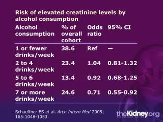
Diet: Protein Restriction • Dietary protein restriction (0.4-0.6 g/kg/day) reduced the risk for renal failure or death in nondiabetic renal disease and slowed nephropathy in type I DM • beneficial effects were unrelated to change in blood pressure or glycemic control. • NNT 5 to 50 • realistic to restrict to 0.8-1g/kg/day Ann Intern Med. 1996 April 1;124: 627-32.
Dyslipidemia • Abnormal lipid metabolism is common in RF • Primary problem is hypertriglyceridemia • LDL, HDL, chol may also be deranged • CRF is considered a high cardiovascular risk and therefore target lipid levels are as those with CAD • Some suggest that statins may benefit the kidney independent of lipid lowering effects
Erythropoietin • Indicated for Hgb <110 • Chk iron stores, folate, and B12 • In patients with ferritin <100 and sat <20%, must supplement prior to commencement of therapy and continue supplement while on EPO • Follow response to EPO and ensure adequate residual stores
Case • Your patient returns for follow-up after 6 months. He has no new symptoms. You repeat his serum creatinine. It is now 185 (from 155). • At what creatinine level would you refer him to a nephrologist? a) Now (185) b) 250 C) 310
Referral to Nephrologist • Late referral (< 12 months pre dialysis) is common • Survey of Ontario Family MDs: • 84% would not refer with creat 120-150 (>50% loss of GFR) • 28% would not refer with creat 150-300 • almost all would refer with creat>300 • Consequences of referral shortly before dialysis: • more complications • longer hospitalization to initiate dialysis • more difficulty with initiation of dialysis • worse survival! • Better outcomes with early multidisciplinary care CMAJ 1999: 161:413-17
Canadian Guidelines • Renal replacement therapy is NOT rationed (i.e. everyone should be considered) • Reversible causes should be sought at diagnosis • At least 1 year is required to prepare for dialysis • Refer, at the latest, at Cr clearance of 30 ml/min, or Cr of 300 • But…there are probably not enough nephrologists/ clinics to meet this demand • Adequate communication with the Nephrologist will allow proper stratification of patients CMAJ 1999: 161:413-17
For AIMGP Clinic • It is reasonable to follow stable renal failure patients, and work up and manage appropriately • Refer to nephrology when: • Cr >300 or Cr clearance <30 ml/min • Renal biopsy indicated • Indicators of aggressive disease are present: • Rapid decline in creatinine • homeostatic derangement i.e. acidosis, volume overload, high K • high protein excretion • Difficult to control BP • low HDL • black race
Conclusions • When evaluating a patient with increased creatinine, internists should: • Identify and treat reversible causes of renal failure • Initiate management to slow the decline in renal function • Manage coexisting conditions • Have clear indications for when to refer to nephrology subspecialists
References • Elevated Serum creatinine: recommendations for management and referral. CMAJ 1999: 161:413-17 • Approach to managing elevated creatinine. Can Fam Physician. 2004;50:735-740. • UptoDate