Download
1 / 21
220 likes | 455 Vues
Anatomy of Bones. From Head to toes to hands. Medial Lateral Anterior Posterior Superior Inferior Proximal Distal Dorsal/dorsum Palmar Plantar Condyle/epicondyle Fossa Process Tuberosity Styloid. Apophysis Diaphysis Epiphysis Medullary cavity Osteoblasts Osteoclasts

E N D
Anatomy of Bones From Head to toes to hands
Medial Lateral Anterior Posterior Superior Inferior Proximal Distal Dorsal/dorsum Palmar Plantar Condyle/epicondyle Fossa Process Tuberosity Styloid Apophysis Diaphysis Epiphysis Medullary cavity Osteoblasts Osteoclasts Periosteum Endoosteum Articular Cartilage/Hyaline Osteocytes Cancellous Bone Wolff’s Law Vocabulary
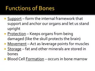
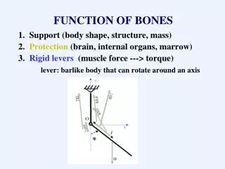
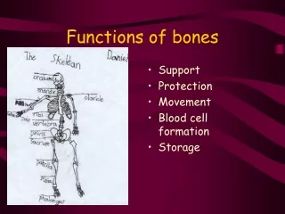
Function of Bone • 1. Body support • 2. Organ protection • 3. Movement (through joints and levers) • 4. Calcium storage • 5. Formation of blood cells (hematopoesis)
Types of Bone • Flat- skull, ribs, scapula • Irregular- vertebral column, skull • Short – wrist and ankle • Long- humerus, ulna, radius, femur, tibia, fibula, phalanges, metqacarpal/metatarsal
How the bone works • You can see: • Diaphysis, epiphysis, articular cartilage, periosteum, medullary cavity, & endosteum • The end of bones have a layer of hyaline cartilage that provide a very smooth surface that protects and cushions blows to the joint. • Periosteum: • Covers long bones • Sharpey’s fibers connect the periosteum to underlying bone • Muscle/tendon fibers interlace the periosteum
Bone Growth • Bone ossification occurs by the synthesis of osteoblasts • Osteoblasts builds new bone on the outside of the bone • Same time osteoclsts increase the medullary cavity by breaking down bony tissue. • Osteogenesis occurs when there is a balance of bone formation and bone destruction
Periostitis Bone fractures Depressed fx Greenstick fracture Impacted fracture Longitudinal fracture Spiral fracture Oblique fracture Serrated fracture Transverse Fx Communited Fx Contrecoup Fx Blowout Fx Avulsion Fx Stress Fx. Epiphyseal Fx: Salter-Harris Type I-V Bone Trauma
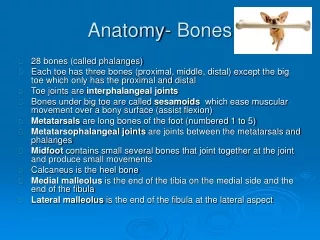
Anatomy Of The Elbow/Wrist/Hand • 1. Navicular (scaphoid) • 2. Lunate • 3. Triquetral • 4. Pisiform • 5. Hamate • 6. Capitate • 7. Trapezoid • 8. Trapezium • 9. Ulnar styloid • 10. Radial Styloid • 11. Distal/middle /proximal phalanx • 12 DIP, PIP, MCP joints
Anatomy Of The Elbow • 1. Humeral Shaft. • 2. Radial Shaft. • 3. Ulnar Shaft. • 4. Olecranon Process. • 5. Head of the Radius. • 6. Coronoid Process of the Ulna. • 7. Medial Supracondylar ridge.
Bones of the elbow 1 Capitulum- 2 Trochlea 3 Lateral epicondyle 4 Medial epicondyle
Radial Shaft. Ulnar Shaft. Humeral Shaft. Medial Epicondyle. Lateral Epicondyle. Radial Tuberosity. Olecranon Process of the Ulna.
Humerus Greater tubercle Lesser tubercle Bicipital groove Head of humerus Scapula Acromion process Coracoid process Glenoid fossa Supra/infraspinatus fossa Subscapularis fossa Superior/inferior border and angle Medial/lateral border Spine of the scapula Proximal Humerus and shoulder region
Superior angle Head of humerus Greater tubercle Supraspinatus fossa Infraspinatus fossa Glenoid fossa Inferior angle Deltoid tuberosity
Shoulder Coracoid process Lesser tubercle Subscapularis fossa Bicipital groove
Clavicle and Sternum • Manubrium • Body of the sternum • Xiphoid Process
Vertebrae Body Vertebral foramen Transverse process facets Spinous process
Vertrebral Column • 7-cervical • 12-thoracic • 5-lumbar • Sacrum • Coccyx • Sacral iliac joint
Hip/pelvis Iliac crest ilium ASIS AIIS ischium Head of femur Greater trochanter ILIAC TUBEROSITY
Hip/pelvis Iliac Crest Iliac Fossa ASIS Greater trochanter Body of Pubis Body of Ischium Lesser trochanter
Knee Lateral femoral condyle Medial femoral condyle Gerdy’s tubercle Fibular styloid Medial tibial condyle Lateral tibial condyle Head of fibula Tibial tuberosity
Posterior knee Popliteal surface Intercondylar fossa Styloid of fibula Posterior iintercondylar fossa