Download
1 / 40
420 likes | 758 Vues
Bones of the. Upper Limb. Skeleton:. The skeleton consists of: Bones : special connective tissue, hard. Cartilage : special connective tissue, less hard than bones.

E N D
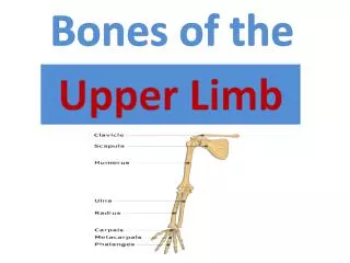
Bones of the Upper Limb
Skeleton: • The skeleton consists of: • Bones: special connective tissue, hard. • Cartilage: special connective tissue, less hard than bones. • Joints: joint is the location at witch two bones make contact, whereas ligaments are the real site of contact (dense proper connective tissue: collagens.) • - Types of joints: fibrous joint, cartilaginous joint, and synovial joint. • Tendons: dense proper connective tissue, connects muscle to bone.
Bones Definition: a calcified connective tissue that stores calcium and phosphorus. Functions: Support and protection. Ex. Skull protects the brain. 2. Serves as a lever, and helps to muscle attachment which is responsible for body movement. Blood cell formation due to the delicate blood forming bone marrow within its cavities. Mineral storage (calcium especially).
Types of Bone Tissue • Classified according to relative amount of solid matrix (Ca\P), number and size of bone marrow cavities: • Compact bone (cortical): • Full with: Ca, P. • 2. Spongy bone (cancellous) (trabecular): • Full with: bone marrow cavities.
Types of Bones Based on Shape: Long bones: two ends (epiphysis) and a shaft (diaphysis). Short bones: cube – shaped bones. Flat bones: thin and curved. Irregular bones: their shapes are irregular and complicated. Sesamoid bones: bones embedded in tendons.
Clavicle • S shaped • Long bone of short length that serves as a strut between scapula and sternum . • The only bone in the body that lies horizontally.
Two ends, two surfaces, and two borders. • Two ends: medial (sternal), and lateral (acromial). • Medial 2/3 is convex, lateral 1/3 is concave. • The acromial end is flat, whereas the sternal end is bulky. • Two surfaces: upper (smooth), and lower (rough), because of the conoid tubercle and the subclavian groove. • Two borders: anterior and posterior.
Common site of fracture is the weak point between medial 2/3 and lateral 1/3 at the junction between two curves. It causes sever pain. • Treatment: attach the weight of the upper limb to the neck.
Functions of clavicle: Connects the axial skeleton with the appendicular. Muscles attachment (origin or insertion), and ligaments attachment. Transmission of weight of the upper limb to the axial skeleton.
Scapula • Flat bone. • Roughly triangular in shape. • Connects the humerus with the clavicle.
Three angles, three borders, two surfaces: • Three angles: superior, inferior, lateral. • Three borders: superior, medial (vertebral), lateral (axillary). • Two surfaces: anterior, posterior.
Spine: prominent plate of bone that separates the suprspinatous from the infraspinatousfossa. • Acromion: flattened lateral portion of the spine of scapula. • Coracoids process: a small hook-like structure on the lateral edge of the superior anterior portion of the scapula. Together with the acromion, serves to stabilize the shoulder joint. It’s palpable. • Glenoid cavity: articulates with the head of humerus. • Suprascapular notch: contains a ligament called suprascapular ligament.
Functions of Scapula: • Muscles and ligaments attachment • Fixation of the shoulder joint • Connection with axial skeleton by clavicle. • Important for movement.
Humerus • Long bone: proximal end, shaft, and distal end.
Head: covered with hyaline cartilage and makes articulation with glenoid cavity. • Anatomical neck: there’s a tendon that comes through it for the biceps muscle. • Surgical neck: treated by surgery. • Intertubercular groove: divided into medial lip, lateral lip, and floor. • Deltoid tuberosity: muscle attachment for deltoid muscle. • Medial condyle (trochlea) is bigger and there’s a nerve cross behind it called “ulnar nerve.” • Olecranonfossa: articulates with olecranon process of ulna.
Glenohumeral Joints (shoulder joint) • -Synovial ball and socket joint between the head of humerus and the glenoid cavity of scapula. • Supports a wide range of movements provided at the cost of skeletal stability. Joint stability is provided, instead, by rotator cuff muscles which blend with the joint capsule to surround the anterior, posterior, and superior aspects of the shoulder joint. • Humerus dislocation is common. • Treatment: surgery, or Kocher’s method.
Ulna and Radius Ulna Radius • The longer of the two forearm bones • Located medially (pinky side) • The head is at the distal end. • Styloid process is located medially. • Notch called radial notch of ulna. • Other structures: olecranon process, trochlear notch, coronoid process. • The shorter of the two forearm bones. • Located laterally (thumb side) • The head is at the proximal end. • Styloid process is located laterally. • Notch called ulnar notch of radius. • Other structures: neck, dorsal radial tubercle.
The two bones are connected by interosseous membrane. • There are proximal and distal radioulnar joints between the two bones: • Proximally: the head of the radius articulates with the radial notch of ulna, forming superior radioulnar joint. • Distally: the head of the ulna articulates with the ulnar notch of radius, forming inferior radioulnar joint.
Elbow Joint • Extension of elbow joint: olecranon process of ulna articulates with the olecranonfossa of humerus. • Flexion of elbow joint: head of radius articulates with radial fossa. Coronoid process of ulna articulates with coronoidfossa of humerus.
Bones of the Hand • Carpals: 16 bones (each hand 8). • Metacarpals: 10 bones (each hand 5). • Phalanges: 28 bones (each hand 14).
Carpal Bones • From lateral to medial: • Proximal row: scaphoid, lunate, triquetrum, pisiform. • Distal row: trapezium, trapezoid, capitate, hamate.
Metacarpal Bones • Numbered from 1 to 5, from lateral to medial. • Base and shaft are concave, head is convex.
Phalanges Two bones in the thumb (proximal and distal), and three in each finger (proximal, middle, and distal).
Wrist Joint • The ulna doesn’t articulate with the carpal bones directly. Instead, there’s a triangular disk between them. • The radius articulates with scaphoid and lunate.
Joints of the Hand • Between carpals: • intercarpal articulations. • Between carpals and metacarpals: • carpometacarpal joints. • Between metacarpals and phalanges: metacarpophalangeal joints. • between phalanges: • interphalangeal articulations.