Download
1 / 12
140 likes | 311 Vues
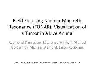
Field Focusing Nuclear Magnetic Resonance (FONAR): Visualization of a Tumor in a Live Animal. Raymond Damadian , Lawrence Minkoff , Michael Goldsmith, Michael Stanford, Jason Koutcher . Dana Braff & Lisa Foo |20.309 Fall 2011| 13 December 2011. Background on R. Damadian.

E N D
Field Focusing Nuclear Magnetic Resonance (FONAR): Visualization of a Tumor in a Live Animal Raymond Damadian, Lawrence Minkoff, Michael Goldsmith, Michael Stanford, Jason Koutcher. Dana Braff & Lisa Foo |20.309 Fall 2011| 13 December 2011
Background on R. Damadian • Inventor of the Magnetic Resonance (MR) scanning machine • Published his 1971 paper “Tumor Detection by Magnetic Resonance” a year prior to filing a patent for a MRI scanner • Formed FONAR Corporation which produced the first commercial scanner in 1980. • Earned the a Lemelson-MIT Program’s Lifetime Achievement Award, National Medal of Technology and was inducted into the National Inventors Hall of Fame.
Brief History of Nuclear Magnetic Resonance • Application in health care was developed in the early 1970’s • Non-invasive technique and safe alternative to X-rays. Findings could be easily and quickly obtained from a single system. • Application of NMR imaging stayed in research and experimental phase through the 70’s and 80’s • Field focusing Nuclear Magnetic Resonance (FONAR) enabled one to externally direct the NMR spot to a anatomic site of interest for continuously monitored inspection.
Nuclear Spin Source: Zimmerman, 20.309 Stellar Website
Precession Source: Zimmerman, 20.309 Stellar Website
NMR: how it works • NMR experiments can be thought of as having two stages: excitation and acquisition • Critical components of NMR: • Large static homogeneous magnetic field, B0 • Coil to generate the resonant excitation field, B1 • A coil used to measure the precession of the spins • A sample that has “spin” property Source: Zimmerman, 20.309 Stellar Website
FONAR • Field Focusing Nuclear Magnetic Resonance • Resonant Window (aperture) • Scanning
Significance • This research was extended towards developing the NMR method as a way of noninvasively detecting chemical abnormalities in human internal organs. • Imaging a human chest • when the exploring n.m.r sample is at the plateau of the conic section a signal is produced. • The signal vanishes when moved off-center along either axis because H0 departs from the value of resonance and because the field is too steeply graded.
Formation of Chemical Images in Man Figure 1. Fonar cross-section of the live human chest at the level of the 8th thoracic vertebra Figure 2. Fonar scan of man with pulmonary oat cell carcinoma. Tumor indicated by blue infiltrate in left lung field. Source: Damadian 1980