Download
1 / 40
510 likes | 1.41k Vues
Sinus Histiocytosis with Massive Lymphoadenopathy (Rosai-Dorfman Disease). Clinical Pathology Conference November 4, 2005 Dean Fong, DO. Disorder of Histiocytic and Dendritic Derivation. Spectrum from benign to frank malignant Problems with diagnosis: Scarcity of specific markers

E N D
Sinus Histiocytosis with Massive Lymphoadenopathy (Rosai-Dorfman Disease) Clinical Pathology Conference November 4, 2005 Dean Fong, DO
Disorder of Histiocytic and Dendritic Derivation • Spectrum from benign to frank malignant • Problems with diagnosis: • Scarcity of specific markers • Lack of consistent means for detection of monoclonality • Clinicopathologic overlap with reactive and infectious proliferations
Non-Malignant Histocytoses • Group of disorders involving a pathologic increase in the number of histiocytes • Mononuclear phagocytic cells • Circulating monocyte • Alveolar macrophages of the lung • Kupffer cells of the liver • Osteoclasts • Microglial cells
Non-Malignant Histocytoses • Mainly-antigen presenting cells • Interdigiting reticulum cells and dendritic reticulum cells in the spleen and lymph nodes • Langerhans cells in skin and bronchial epithelium • Bone marrow origin
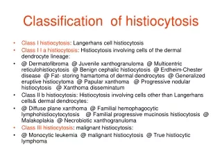
Non-Malignant Histocytoses • Three group of disease • Dendritic cell-related histiocytoses • Langerhans cell histiocytoses • Histiocytosis X • Eosinophilic granuloma • Hand-Schuller-Christian disease • Letterer-Siwe disease • Single system disease • Multisystem disease • Juvenile xanthogranuloma-dermal dendrocyte phenotype
Non-Malignant Histocytoses • Three group of disease (cont.): • Macrophage-related histiocytoses • Hemophagocytic Lymphohistiocytosis • Primary hemophagocytic lymphohistiocytosis or familial hemophagocytic lymphohistiocytosis • Sporadic or familial • Associated with infection • Secondary hemophagocytic lymphohistiocytosis • Infection-associated hemophagocytic syndrome • Malignancy associated hemophagocytic syndrome • Others, including fat overload syndrome • Rosai-Dorfman disease
Sinus Histiocytosis with Massive Lymphoadenopathy (SHML) • First described by Rosai and Dorfman in 1969. • Nonmalignant proliferation of distinctive histiocytic/phagocytic cells within lymph node sinuses and lymphatics in extranodal sites
Sinus Histiocytosis with Massive Lymphoadenopathy (SHML) • Clinical features • Worldwide • Primarily disease of childhood and early adulthood • Peak age 20 years • Increased incidence of serum auto-immune antibodies during active disease • No specific gender, ethnic, or socioeconomic predilection • Some reports of M > F
Sinus Histiocytosis with Massive Lymphoadenopathy (SHML) • Clinical features • Registry of 423 cases: • Caucasian = African • Asian Less common • Occasional familial cases
Pathogenetic Mechanism • Early 3 of 6 cases found serologic evidence of EBV • In 7 of 9 pts. HHV-6 DNA found • Unfavorable outcome in patients with immune dysfunction • Exuberant response of hematopoietic system to undetermined immunologic trigger • ? Defective Fas/FasL signaling leading to defective apoptosis ? histiocytic proliferation
Sinus Histiocytosis with Massive Lymphoadenopathy (SHML) • Most frequent presenting symptoms • Cervical region painless lymphadenopathy • Up to 90% of cases • Axillary, para-aortic, inguinal and mediastinal lymph nodes are commonly affected • Extranodal disease in 43% of patients
Skin Involvement Firm indurated papules
Sinus Histiocytosis with Massive Lymphoadenopathy (SHML) • Antecedent non-specific fevers and pharyngitis may herald the onset of SHML • Occasionally accompanied by pain, tenderness, malaise, night sweats or weight loss
Pathological Features • Laboratory findings: • Normocytic or microcytic anemia • Immunologic abnormalities significant number of pts. unfavorable prgnosis • 90% pts. elevated ESR • Most frequent immune dysfunction AIHA • Polyarthralgia, RA, glomerulopathies, asthma, DM complicate SHML • Polyclonal hypergammaglobinemia 90% of pts. • Rare RF, ANA, reversal of CD4/CD8 • Small subset NHL, other histiocytic proliferations, myeloma, melanoma, CA • Reported EBV and HHV-6
Pathology • Gross • Yellow-white with frequent capsular and pericapsular fibrosis
Microscopic • Normal lymph node architecture preserved • Effacement seen only in pts. with long-standing lymphadenopathy • Lymph node sinuses expanded by proliferation of distinctive histiocytes
Histiocytes • Enlarged round or oval vesicular nuclei with well defined, delicate nuclear membranes and a single prominent nucleolus • Multilobulated nuclei, nucleus with multiple nucleoli, nuclear atypia rare • Mitoses infrequent but increased mitotic activity can be apparent occasionally • Abundant pale eosinophilic cytoplasm • Occasional numerous histiocytes with foamy cytoplasm may predominat cellular milieu
Histiocytes • Hallmark lymphophagocytosis or emperipolesis • Lymphocytic penetration and movement within another cell • Often housed within vacuoles escape degradation • Plasma cells, PMNs, RBCs may also be present
Other Histopathological Features • Plasma cells often aggregated around post-capillary venules • Eosinophils not usually seen if seen, think: • LCH, HL, T-cell lymphoma • Collections of PMNs, eosinophilic microabscess, reactive germinal centers seen but not prominent features • Extranodal sites more fibrosis, and fewer histiocytes with emperipolesis
Differential Diagnosis • Langerhans Cell Histiocytosis • Lymph node sinuses expanded by histiocytes seen in both LCH and SHML but… • LCH cells are frequently folded or grooved nuclei and associated with eosinophilic microabscess • Histocytic sarcoma • Storage disease • Gaucher’s disease • Hodgkin Lymphoma
Differential Diagnosis • Metastatic melanoma • Carcinoma • Infections caused by: • Histoplasma • Mycobacterial organism • Reactive sinus histiocytosis
Differential Diagnosis • Emperipolesis rare outside setting of SHML but is seen in reactive, neoplastic histiocytic proliferation, LCH
Immunohistiologic Studies • Most useful immunologic marker histiocytes with expression of S100 • Histiocytes • Pan-macrophages antigens CD68, HAM 56, CD14, CD64, CD15 • Antigens associated with phagocytosis CD64, Fc receptor for IgG • Lysosomal activity Lysozyme, A1A • Immune activation Transfering receptor, IL-2 receptor • CD163 hemoglobin scavenger receptor and acute phase-regulated transmembrane protein found on tissue macrophages and monocytes
Immunohistiologic Studies • Effector cells in SHML • Functionally activated macrophages • Distinct from Langerhans cells, follicular dendritic cells, interdigiting dendritic cells
Clinical course and treatment • Characterized by spontaneous resolution in most cases • Usually indolent for many years, with spontaneous regression • Do not usually threaten life or organ function • Few pts. disease progressive and require treatment • Some pts. episodes of exacerbation alternating with periods of remission that continue for many years
Clinical course and treatment • Persistent lymphadenopathy or progression • Associated with involvement of the kidney, lower respiratory tract or liver with associated immunologic dysfunction • Poor prognosis
Clinical course and treatment • SHML registry 423 cases 17 deaths • Only few pts. warrant treatment no randomized trials • “Wait-and-see” approach • Antibiotics or anti-tuberculosis drugs no response • Steroids reduction in lymphoadenopathy and associated fevers • Associated autoimmune conditions usually resolve as the primary condition responds to steroid therapy
Clinical course and treatment • Radiation • 3 complete remission • 3 persistent SHML • 3 death • Chemotherapy • 10 no response • 2 complete and durable remission • Surgery and radiation • 1 complete remission • 6 partial remission • High dose interferon α long-term remission • No ideal treatment more data needed
Late Sequelae and Follow-Up • Few pts. require prolonged or intermittent treatment with corticosteroids • Long term steroid effects • No increased incidence of secondary tumors • Follow-up • Monitor disease with clinical examination and CXR
References • Henter JI, Tondini C et. al., “Histiocyte disorders”, Critical Reviews in Oncology Hematology, 2004; 50: 157-174. • Mills SE et. al., Sternberg’s Diagnostic Surgical Pathology, 4th Ed., 2004; 479. • McClain KL, Natkunam Y, et. al., “Atypical Cellular Disorders”, Hematology 2004. • Weitzmann S, Jaffe F, “Uncommon Histiocytic Disorders: The Non-Langerhans Cell Histiocytosis”, Pediatr Blood Cancer, 2005; 45: 256-264.