Download
1 / 6
60 likes | 69 Vues
The anesthetic problems during minimal access surgery are related to the cardiopulmonary effects of pneumoperitoneum, carbon dioxide (CO2) absorption, extraperitoneal gas insufflation, venous embolism, and inadvertent injuries to intraabdominal organs.

E N D
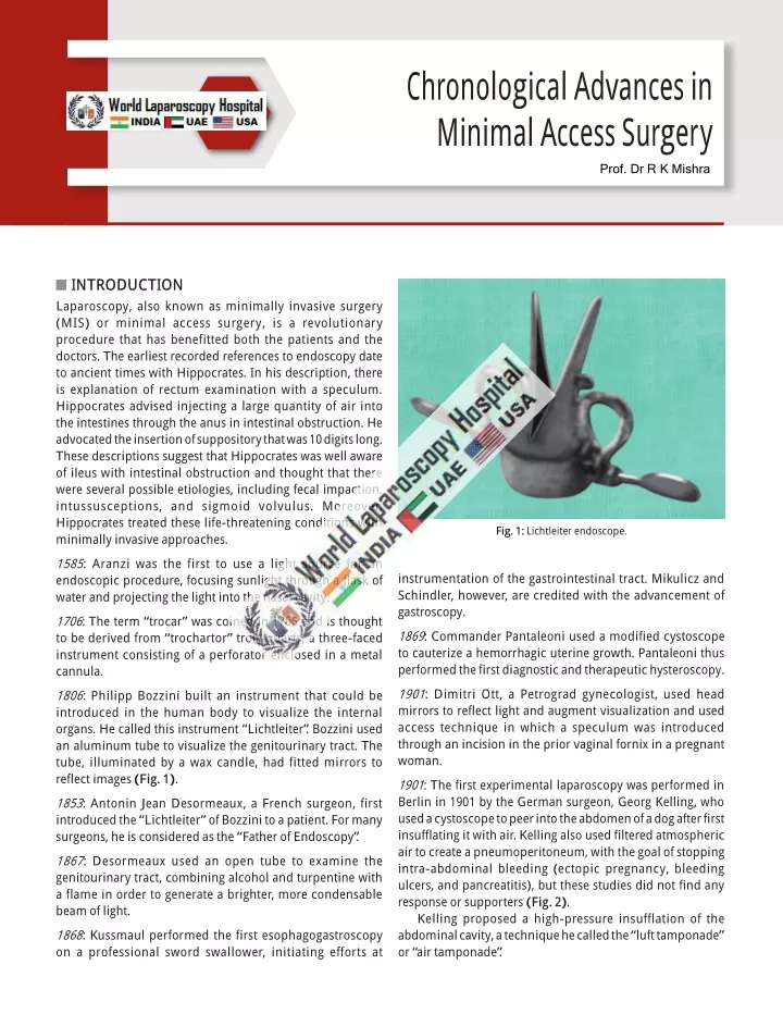
C hronological Advances in M inim al Access Surgery Prof. Dr R K Mishra INTRODUCTION Laparoscopy, also known as minimally invasive surgery (MIS) or minimal access surgery, is a revolutionary procedure that has benefitted both the patients and the doctors. The earliest recorded references to endoscopy date to ancient times with Hippocrates. In his description, there is explanation of rectum examination with a speculum. Hippocrates advised injecting a large quantity of air into the intestines through the anus in intestinal obstruction. He advocated the insertion of suppository that was 10 digits long. These descriptions suggest that Hippocrates was well aware of ileus with intestinal obstruction and thought that there were several possible etiologies, including fecal impaction, intussusceptions, and sigmoid volvulus. Moreover, Hippocrates treated these life-threatening conditions with minimally invasive approaches. Fig. 1: Lichtleiter endoscope. 1585: Aranzi was the first to use a light source for an endoscopic procedure, focusing sunlight through a flask of water and projecting the light into the nasal cavity. instrumentation of the gastrointestinal tract. Mikulicz and Schindler, however, are credited with the advancement of gastroscopy. 1706: The term “trocar” was coined in 1706 and is thought to be derived from “trochartor” trois-quarts, a three-faced instrument consisting of a perforator enclosed in a metal cannula. 1869: Commander Pantaleoni used a modified cystoscope to cauterize a hemorrhagic uterine growth. Pantaleoni thus performed the first diagnostic and therapeutic hysteroscopy. 1901: Dimitri Ott, a Petrograd gynecologist, used head mirrors to reflect light and augment visualization and used access technique in which a speculum was introduced through an incision in the prior vaginal fornix in a pregnant woman. 1806: Philipp Bozzini built an instrument that could be introduced in the human body to visualize the internal organs. He called this instrument “Lichtleiter”. Bozzini used an aluminum tube to visualize the genitourinary tract. The tube, illuminated by a wax candle, had fitted mirrors to reflect images (Fig. 1). 1901: The first experimental laparoscopy was performed in Berlin in 1901 by the German surgeon, Georg Kelling, who used a cystoscope to peer into the abdomen of a dog after first insufflating it with air. Kelling also used filtered atmospheric air to create a pneumoperitoneum, with the goal of stopping intra-abdominal bleeding (ectopic pregnancy, bleeding ulcers, and pancreatitis), but these studies did not find any response or supporters (Fig. 2). Kelling proposed a high-pressure insufflation of the abdominal cavity, a technique he called the “luft tamponade” or “air tamponade”. 1853: Antonin Jean Desormeaux, a French surgeon, first introduced the “Lichtleiter” of Bozzini to a patient. For many surgeons, he is considered as the “Father of Endoscopy”. 1867: Desormeaux used an open tube to examine the genitourinary tract, combining alcohol and turpentine with a flame in order to generate a brighter, more condensable beam of light. 1868: Kussmaul performed the first esophagogastroscopy on a professional sword swallower, initiating efforts at
4 SECTION1: Essentials of Laparoscopy Fig. 2: Kelling performing laparoscopy in dog. Fig. 3: Heinz Kalk. 1910: HC Jacobaeus of Stockholm published a paper on discussion of the inspection of the peritoneal, pleural, and pericardial cavity. 1911: Bertram M Bernheim of Johns Hopkins Hospital introduced first laparoscopic surgery to the United States. He named it the procedure of minimal access surgery as “organoscopy”. The instrument used was a proctoscope of a half inch diameter and ordinary light for illumination was used. 1911: HC Jacobaeus coined the term “laparothorakoskopie” after using this procedure on the thorax and abdomen. He used to introduce the trocar inside the body cavity directly without employing a pneumoperitoneum. 1918: O Goetze developed an automatic pneumo peri- toneum needle characterized for its safe introduction to the peritoneal cavity. The next decade and a half witnessed a decline in technological advancement in endoscopy due to World War I. Fig. 4: John C Ruddock. superior than laparotomy. He used the instrument for diagnostic laparoscopy which consisted of built-in forceps with electrocoagulation capacity (Fig. 4). 1936: Boesch of Switzerland is credited with the first laparoscopic tubal sterilization. 1920: Zollikofer of Switzerland discovered the benefit of carbon dioxide (CO2) gas to use for insufflation rather than filtered atmospheric air or nitrogen. 1938: Janos Veress of Hungary developed an especially designed spring-loaded needle. Interestingly, he did not promote the use of his Veress needle for laparoscopy purposes. He used Veress needle for the induction of pneumothorax. Veress needle is widely used instrument today to create pneumoperitoneum (Fig. 5). 1929: Kalk, a German physician, introduced the forward oblique (135°) view lens systems. He advocated the use of a separate puncture site for pneumoperitoneum. Goetze of Germany first developed a needle for insufflation. 1929: Heinz Kalk, a German gastroenterologist, developed a 135° lens system and a dual trocar approach. He used laparoscopy as a diagnostic method for liver and gallbladder diseases (Fig. 3). 1939: Richard W Te Linde tried to perform an endoscopic procedure by a culdoscopic approach in the lithotomy position. This method was rapidly abandoned because of the presence of small intestine. 1939: Heinz Kalk published his experience of over 2,000 liver biopsies performed using local anesthesia without mortality. 1934: John C Ruddock, an American surgeon, described laparoscopy as a good diagnostic method, many times,
5 CHAPTER1: Chronological Advances in Minimal Access Surgery Fig. 5: Veress needle. Fig. 6: Laparoscope. Fig. 7: Kurt Semm. Fig. 8: First insufflator made by Professor Semm. 1944: Raoul Palmer of Paris performed gynecological examinations using laparoscopy (Fig. 6) and placing the patients in the Trendelenburg position, so air could fill the pelvis. He also stressed the importance of continuous intra- abdominal pressure monitoring during a laparoscopic procedure. in his own country, but on the other side of the Atlantic, both American physicians and instrument makers valued the Semm’s insufflator for its simple application, clinical value, and safety (Fig. 8). 1960: Patrick Steptoe, a British gynecologist, adapted the techniques of sterilization by two puncture technique. 1953: The rigid rod lens system was discovered by Professor Hopkins. The credit of videoscopic surgery goes to this surgeon who had revolutionized the concept by making this instrument. 1972: H Courtenay Clarke showed laparoscopic suturing technique for hemostasis. 1973: Gaylord D Alexander developed techniques of safe local and general anesthesia suitable for laparoscopy. 1960: Kurt Semm was a German gynecologist, who invented the automatic insufflator (Fig. 7). His experience with this new device was published in 1966. Apart from insufflator, he also perfected many technical refinements including suction irrigator, safer electrocoagulation instruments, intracorporeal and extracorporeal knot tying, and an electrical morcellator for myomas. Though not recognized 1977: First laparoscopic-assisted appendicectomy was performed by Dekok. Appendix was exteriorized and ligated outside the abdominal cavity. 1977: Kurt Semm demonstrated endoloop suturing technique in laparoscopic surgery.
6 SECTION1: Essentials of Laparoscopy 1978: Hasson introduced an alternative method of blunt trocar placement. He proposed a blunt minilaparotomy which permits direct visualization of trocar entrance into the peritoneal cavity. Hasson cannula was a reusable device of similar design to a standard cannula, but attached to an olive-shaped sleeve was developed. This sleeve would slide up and down over the shaft of the cannula and form an airtight seal at the fascial opening. In addition, the sharp trocar was replaced by a blunt obturator. This cannula is held in place by the use of stay sutures passed through the fascial edges and attached to the body of the cannula (Fig. 9). 1987: Ger reported first laparoscopic repair of inguinal hernia using prototype stapler. 1988: Harry Reich performed laparoscopic lympha- denectomy for treatment of ovarian cancer. 1988: McKernan and Sye performed first cholecystectomy in the USA (Fig. 11). 1989: Harry Reich described first laparoscopic hysterectomy using bipolar desiccation; later, he demonstrated staples and finally sutures for laparoscopic hysterectomy. 1989: Reddick and Olsen reported that common bile duct (CBD) injury after laparoscopic cholecystectomy is five times than with conventional cholecystectomy. As a result of this report, USA government announced that surgeons should perform at least 15 laparoscopic cholecystectomy under supervision before being allowed to do this procedure on their own. 1980: Patrick Steptoe started to perform laparoscopic procedures first time in UK. 1983: Kurt Semm, a German gynecologist, performed the first laparoscopic appendicectomy. After this surgery, Kurt Semm faced a lot of criticism and censor rather than accolades. The German Board of Surgery condemned him. 1990: Bailey and Zucker in USA popularized laparoscopic anterior highly selective vagotomy combined with pos- terior truncal vagotomy. Over the next few years, virtually every abdominal operation was performed and perfected by laparoscopic technique. Laparoscopic splenectomy was performed by Makoto Hashizume, Ed Phillips, Joseph Petelin and John Flowers, and adrenalectomy by Petelin and Gagner. 1985: The first documented laparoscopic cholecystectomy was performed by Erich Mühe in Germany in 1985. The German surgeon Erich Mühe used his “galloscope”, a 3-cm, direct-vision laparoscope of his own design to remove a gallbladder. He presented his work at the 1986, Congress of the German Surgical Society. He, too, suffered skepticism and criticism and was ultimately censored by the courts. 1987: Philippe Mouret has got the credit to perform the first laparoscopic cholecystectomy in Lyons, France using video technique (Fig. 10). Cholecystectomy is the laparoscopic procedure which revolutionized the general surgery. Like Mühe and Semm, Mouret was also strongly criticized. François Dubois, another Frenchman, was the second surgeon to perform videolaparoscopic cholecystectomy in 1988 and was the first to publish his early experience. 1994: First robotic arm was designed to hold the telescope with the goal of improving safety and reducing the need of skilled camera operator (Fig. 12). 1996: First live telecast of laparoscopic surgery performed remotely via the internet (robotic telesurgery). 1997: Fully laparoscopic aortofemoral bypass was being performed, led by Dion and Gracia. Fig. 9: Hasson cannula. Fig. 10: Philippe Mouret.
7 CHAPTER1: Chronological Advances in Minimal Access Surgery Fig. 11: William Sye. Fig. 12: Robotic arm. 2000: The US Food and Drug Administration (FDA) first time approved the da Vinci Surgical System, making it the first robotic system allowed to be used in American operating rooms. Recently, SILS and robotic surgery have penetrated all specialties of abdominal surgery. However, evidence-based medicine has failed to show major advantages in SILS and the disadvantage of robotic surgery is the high costs related to purchase and maintenance of technology. 2001: The Lindbergh operation, named in honor of American aviator Charles Lindbergh, was the first ever transatlantic surgery. Doctors Michel Gagner and Jacques Marescaux removed the gallbladder of a 68-year-old woman in Strasbourg, France from New York. The surgeons used a ZEUS Robotic Surgical System from Computer Motion, Inc., and an ATM fiberoptic connection provided by France Telecom. 2014: Leonard et al. provided a proof-of-concept Smart Tissue Anastomosis Robot (STAR) which was a vision-guided robotic system for laparoscopic suturing. STARs architecture and interface allowed surgeons to manually select and track incisions and the placement of stitches or automatically performed equally spaced stitches based on the contour of the incision. 2004: Robotic prostatectomy became the first most commonly performed robotic surgery. According to Intuitive Surgical, Inc., the California-based manufacturers of the da Vinci Robot, the number of robotic prostatec tomies rose from 36 in 2000 to 8,000 in 2004. 2014: The FDA has announced a series of actions aimed at limiting the use of power morcellators in gynecologic surgeries in April 2014. Recommendations are that morcellator should only be used in certain laparoscopic procedures and that they always be used with equipment designed to contain the spread of any minced-up tissue. 2005: Combining robotically-assisted coronary artery bypass surgery (CABG) with stented angioplasty shows promise for treating extensive coronary artery disease, researchers reported at the American Heart Association Scientific Sessions 2005, Dallas. The evolution of minimal access therapy aims to minimize the traumatic insult to the patient without compromising the safety and efficacy of the treatment compared with traditional open surgery. If this is achieved, patients recover more quickly, which reduces hospital stay and allows more rapid return to full activity and work within minimum time. This year of 2005 was also known as start of first single-incision laparoscopic surgery (SILS). The evolution of MIS has led to the development of laparoendoscopic single-site surgery (LESS) or SILS. SILS was first performed for the treatment of appendicitis at the Department of Pediatric Surgery, Dokuz Eylul Medical School, Izmir, Turkey. 2017: Prior to February 2017, laparoscopic cholecystectomy was performed without routine intraoperative imaging of the biliary tree. Beginning February 2017, laparoscopic cholecystectomy was performed with indocyanine green (ICG). 0.5 mL of ICG was given via intravenous infusion 45 minutes prior to surgery. Fluorescence imaging was performed using the PINPOINT endoscopic fluorescence imaging system (Novadaq, Canada). The triangle of Calot was exposed to display the biliary tree. This is followed by dissection of the triangle of Calot to expose the cystic duct and artery. The cystic artery can be independently visualized a few minutes following ICG injection. Hereafter, both structures are safely clipped and divided. 2020: TransEnterix received the FDA clearance to add an artificial intelligence feature to its Senhance Surgical Robotic System (Fig. 13). The new Intelligent Surgical Unit on the
8 SECTION1: Essentials of Laparoscopy 10. Jacobeus HC. Ueberdie Möglichkeitdie Zystoskopiebei Untersuchungseröser Höhlungenanzuwenden. Münch Med Wochenschr. 1910;57:2090-2. 11. Jacobs M, Verdeja JC, Goldstein HS. Minimally invasive colon resection (laparoscopic colectomy). Surg Laparosc Endosc. 1991;1:133-50. 12. Kalk H. Erfahrungenmitder Laparoskopie (Zugleichmit Beschreibunge inesneuen Instrumentes). Zeitschr Klin Med. 1929;111:303-48. 13. Kelling G. Ueber Oesophagoskopie, Gastroskopieund Kölioskopie. Münch Med Wochenschr. 1902;49:21-4. 14. Kurze W. Uebersichtübermeine Erfahrungenmitder Laparo- thoraskopie. Münch Med Wochenschr. 1911;58:2017-9. 15. Lau WY, Leow CK, Li AK. History of endoscopic and laparo scopic surgery. World J Surg. 1997;21:444-53. 16. Mühe B. The first laparoscopic cholecystectomy. Langenbecks Arch Chir. 1986;369:804. 17. Nadeau OE, Kampmeier OF. Endoscopy of the abdomen: abdominoscopy. Surg Gynecol Obstet. 1925;41:259-71. 18. Nitze M. Beobachtungs- und Untersuchungsmethodefür Harnröhre, Harnblase and Rektum. Wiener Mediz Wochenschr. 1879;29:651-2. 19. Orndorff BH. The peritoneoscope in diagnosis of diseases of the abdomen. J Radiol. 1920;1:307-25. 20. Roccavilla A. L’endoscopia delle grandi cavita sierose mediante un nuovo apparecchio ad illuminazione dirtta (laparo-toracoscopia diretta). La Riforma Medica. 1914;30:991-5. 21. Rosin D. History. In: Rosin D (Ed). Minimal Access Medicine and Surgery. Oxford: Radcliffe Medical Press; 1993. 22. Ruddock JC. Peritoneoscopy. Surg Gynecol Obstet. 1937; 65:623-39. 23. Semm K. Endoscopic appendectomy. Endoscopy. 1983;15:59-64. 24. Semm K. Operative Manual for Endoscopic Abdominal Surgery. Chicago: Thieme Medical Publishers; 1987. 25. Semm K. The history of endoscopy. In: Vitale GC, Sanfilippo JS, Perissat J (Eds). Laparoscopic Surgery: An Atlas for General Surgeons. Philadelphia: Lippincott Williams and Wilkins; 1995. 26. Short AR. The uses of coelioscopy. Br Med J. 1925;3:254-5. 27. Steiner OP. Abdominoscopy. Surg Gynecol Obstet. 1924; 38:266-9. 28. Stone WE. Intra-abdominal examination by the aid of the peritoneoscope. J Kan Med Soc. 1924;24:63-6. 29. Veress J. Neues Instrument zur Ausführung von Brust-oder Bauchpunktionen and Pneumothoraxbehandlung. Deutsch Med Wochenschr. 1938;40:1480-1. 30. Vitale GC, Cuschieri A, Perissat J. Guidelines for the future. In: Vitale GC, Sanfilippo JS, Perissat J (Eds). Laparoscopic Surgery: An Atlas for General Surgeons. Philadelphia: Lippincott Williams and Wilkins; 1995. 31. Zollikofer R. Zur Laparoskopie. Schweiz Med Wochenschr. 1924;5:264-5. Fig. 13: Senhance Surgical Robotic System. system enables the camera to automatically move for a surgeon during a procedure, responding to commands, and recognizing certain objects and locations in the surgical field. BIBLIOGRAPHY 1. Bernheim BM. Organoscopy: cystoscopy of the abdominal cavity. Ann Surg. 1911;53:764-7. 2. Bozzini P. Lichtleiter, eine Erfindung zur Anschau-ung innerer Theile und Krankheiten. J Prakt Arzneikunde. 1806;24:107-13. 3. Desormeaux AJ. The Endoscope, and its Application to the Diagnosis and Treatment of Affections of the Genito-urinary Passages: Lessons Given at Necker Hospital. Chicago: Robert Fergus’ Sons Printers; 1867. 4. Fervers C. Die Laparoskopie mit dem Cystoskop. Mediz Klinik. 1933;31:1042-5. 5. Fourestier M, Gladu A, Vulmiere J. Perfection nements al’ endoscopie medicale. Realisation bronchoscopique. La Presse Medicale. 1952;60:1292-3. 6. Goetze O. Die Röntgendiagnostik bei gasgefüllten Bauchhöhle. Eine neue Methode. Münch Med Wochenschr. 1918;65:1275-80. 7. Gordon AG, Magos AL. The development of laparoscopic surgery. Baillieres Clin Obstet Gynaecol. 1989;3:429-49. 8. Gow JG, Hopkins HH, Wallace DM. The modern urological endoscope. In: Hopkins HH (Ed). Handbook of Urological Endoscopy. Edinburgh: Churchill Livingstone; 1978. 9. Gunning JE. The history of laparoscopy. J Reprod Med. 1974;12:222-5.