Download
1 / 24
240 likes | 278 Vues
Explore the intricate details of the spinal cord, from its anatomical structure to its functional segments and neurological significance. Learn about the arterial supply, meninges, and components that make up this vital part of the central nervous system.

E N D
DEMO – I(Spinal Cord) Ali JassimAlhashli Year IV – Unit VIII - CNS
STATION – 1 (Gross Anatomy) • The central nervous system (CNS) is composed of: • Brain: which is further composed of: • Forebrain: containing the cerebral hemispheres and diencephalon. • Midbrain. • Hindbrain: containing pons, medulla and cerebellum. • Spinal cord. • The brainstem is composed of 3 parts (see the image): • Midbrain. • Pons. • Medulla. • The spinal cord: • It is the continuation of medulla oblongata extending from foramen magnum to: • Level of IV disc between L1-L2 → in adults. • Level if L3 vertebra → in children. • As you descend, you will notice that the vertebral column is longer than the spinal cord → thus the nerve roots of lower lumbar and sacral regions will descend further down as bundles of nerve roots known as caudaequina.
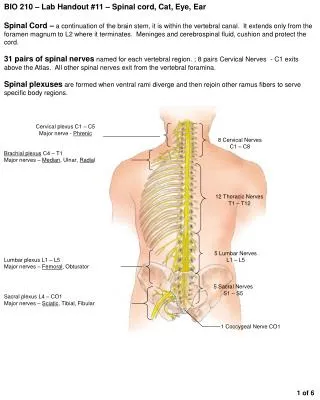
STATION – 1 (Gross Anatomy) • The spinal cord is divided into imaginary segments related to vertebral bones. These segments are: • 8 cervical → corresponding to 7 cervical vertebrae. • 12 thoracic → corresponding to 12 thoracic vertebrae. • 5 lumbar → corresponding to 5 lumbar vertebrae. • 5 sacral → corresponding to 5 sacral vertebrae. • 1 coccygeal→ corresponding to 4 cocygeal vertebrae. • The spinal cord has a ventral horn (motor function) and a dorsal horn (sensory function). The ventral and dorsal nerve roots will merge together to form the spinal nerve. The spinal nerve will then divide into anterior and posterior rami. • There are 31 pairs of spinal nerves exiting the vertebral canal through intervertebral foramina. • C1-C7 spinal nerves will exit above their corresponding vertebrae. C8 spinal nerve will exit above T1 vertebra (because there is no C8 vertebra). Therefore, the rest of spinal nerves will exit below their corresponding vertebrae.
STATION – 1 (Gross Anatomy) • Vertebrae and corresponding spinal cord segments: • Remember that the origin of phrenic nerve is from: C3, C4, C5. if there is an injury to the spinal cord above C4 → quadriplagia will result (paralysis of all limbs) in addition to repiratory deficit. • If there is a lesion to the spinal cord below T1 → paraplagia will result (paralysis of both lower limbs while upper limbs will be spared). • If the lesion is at the level of L2 or below → the spinal cord will not be affected because it ends at the level of IV disc between L1-L2 but the caudaequina (nerve roots) will be injured.
STATION – 1 (Gross Anatomy) • Arterial supply of the spinal cord: the spinal cord is supplied by 3 small arteries: • Anterior spinal artery: • Running in the ventral fissure of the spinal cord. • Formed by merging branches from vertebral arteries. • Supplying the anterior 2/3 of the spinal cord. • 2 posterior spinal arteries: • Formed by branches from vertebral arteries or posterior inferior cerebellar arteries. • Note: radicular (feeder) arteries enter via intervertberal foramina and reinforce anterior and posterior spinal arteries and supply the dorsal root ganglia. Radicular arteries are coming from: • Cervical arteries: in the cervical region. • Intercostal arteries: in the thoracic region. • Lumbar arteries: in the lumbar region.
STATION – 1 (Gross Anatomy) • Meninges (see image in next slide): • Pia matter: it is the innermost covering of the spinal cord which is directly attached to it. It extends as filumterminale which is attached to the 1stcocygeal vertebra. There are extensions from the pia mater going to the dura mater known as denticulate ligaments. • Dura matter: it is the outermost covering of the spinal cord which is extending to the level of S2 vertebra. It is lining the vertebral canal. • Arachenoid matter: it is the internal lining of the dura. The subarachenoid space contains the cerebro-spinal fluid (CSF) and cerebral vessels. • Lumbar puncture: • It is done at the level of: • L3-L4 in adults. • L4-L5 in children. • The structures which will be passed by the needle during lumbar puncture are: • Skin and subcutaneous tissue. • Supraspinous ligament. • Interspinous ligament. • Ligamentumflavum. • epidural space. • Dura. • Subdural space. • Arachenoid. • Subarachnoid space.
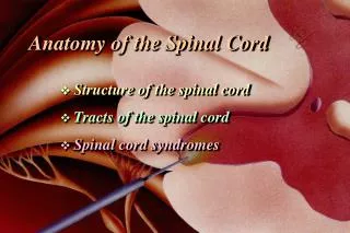
STATION – 2 (Components of Spinal Cord) • The spinal cord has a: • Dorsal median sulcus. • Ventral median fissue. • The spinal cord is composed of: • Grey mater: which is H-shaped or butterfly-shaped and appearing grey because it is containing cell bodies (neurons) with nuclei and different organelles. It is divided into 2 major parts: • The ventral horn: which is concerned with motor function and containing lower motor neurons (α-motor neurons). • The dorsal horn: which is concerned with sensory function. • White mater: appearing white because of lipid of myelin sheath which is surrounding the axons. It is represented by: • The dorsal column: which has mainly the dorsal column medial lemniscus tract. • 2 lateral column: which have mainly lateral spino-thalamic tract + ventral and dorsal spinocerebellar tracts. • The anterior column: which has mainly the anterior spino-thalamic tract.
STATION – 2 (Components of Spinal Cord) • Myelin sheath: it is produced by: • Schwann cells: in peripheral nervous system (PNS). • Oligodendrocytes: in central nervous system (CNS). • As you descend through the spinal cord (from cervical segments down to the thoracic and lumbar segments), you will notice that: • The ventral horn of the cervical and lumbar segments is big → because it is supplying upper and lower limbs (through brachial, lumbar and sacral plexuses). • The ventral horn of the thoracic segment is small → because it is only supplying axial muscles. • A lateral horn is present in the thoracic segments → this is concerned with motor activity of the viscera. • White mater increases as you ascend toward cervical segments → because all ascending tracts are present and merge at this point.
STATION – 2 (Components of Spinal Cord) • Ascending tracts of the spinal cord: • Dorsal column-ML pathway: transmitting sensations of fine touch, vibration, conscious proprioception and 2-point discrimination. • Spinothalamic tract: • Lateral spinothalamic tract: transmitting sensations of pain and temperature. • Anterior spinothalamic tract: transmitting crude touch sensation. • Spinocerebellar tract: for unconscious proprioception. • Classification of sensory receptors: • Vibration: pacinian. • Temperature: thermoreceptors. • Pain: free nerve endings. • Proprioception: muscle spindles, Golgi tendon organs and joint-receptors. • Visceral receptors: chemoreceptors, baroreceptors and osmoreceptors.
STATION – 2 (Components of Spinal Cord) • Dorsal column – ML pathway: • 1st order neurons: sensory fibers enter the spinal cord through the dorsal horn and ascend directly (without synapse) in the dorsal column as f.gracilis and f.cuneatus to terminate in n.gracilis and n.cuneatus of the medulla. • 2nd order neurons: from n.gracilis and n.cunetus, fibers will cross (forming internal arcuate fibers) and ascend as medial lemniscus to terminate in the VPL nucleus of the thalamus. • 3rd order neurons: from VPL nucleus of the thalamus to the post-central gyrus (sensory) of the cerebral cortex. • Spinothalamic tract: • 1st order neurons: sensory fibers enter the spinal cord through the dorsal horn and ascend 2-3 segments to synapse and cross the midline. • 2nd order neurons: they will ascend as spinal lemniscus to terminate in the VPL nucleus of the thalamus. • 3rd order neurons: from VPL nucleus of the thalamus to post-central gyrus (sensory) of the cerebral cortex. • Spinocerebellar pathway (transmitting information of unconscious proprioception): • Dorsal: fibers entering the spinal cord through the dorsal horn and synapse at the level of entry (with Clark’s nuclei) and then ascend ipsilaterally to terminate in the cerebellum through the inferior cerebellar peduncle. • Ventral: fibers entering the spinal cord through the dorsal horn, they syanpse and cross the midline at the level of entry then ascend from the opposite side to recross and terminate in the cerebellum through the superior cerebellar peduncle.
STATION – 2 (Components of Spinal Cord) • Descending tracts of the spinal cord: they are divided into: • Pyramidal: • Cortciospinal tract (movement of the body): descending from the primary motor cortex to lower motor neurons in the spinal cord. fibers are passing through cruscreberi (in midbrain), dispersed (in pons), passing through pyramids (in medulla) and then (90%) of fibers will cross in lower medulla to reach the spinal cord as lateral corticospinal tract while (10%) of fibers will not cross and descend to spinal cord as anterior corticospinal tract. • Corticonuclear tract (movement of the face): descending fibers from the cortex to nuclei of cranial nerves in the brainstem. • Extrapyramidal: • Tectospinal: mediating visual-spinal reflex. Fibers originating from supriorcolliculus in the midbrain. • Vestibulospinal: activating extensor muscles. • Rubrospinal: activating flexor muscles. Fibers originating from red nucleus in the midbrain. • Reticulospinal: • Pontine reticulospinal: helping vestibulospinal in contracting extensors. • Medullaryreticulospinal: helping rubrospinal in contracting felxors.
STATION – 3 (Embryology) • Development of the neural tube: • The notochord induces the overlying ectoderm to differentiate into neuroectoderm and form the neural plate. • The neural plate folds to give rise to the neural tube which is open at both ends at the anterior and posterior neuropores. • These neuropores will close at the 1st month of development. • The lumen of the neural tube gives rise to the ventricular system of the brain and central canal of the spinal cord.
STATION – 3 (Embryology) • Parts of the brain: • Forebrain = prosencephalon. Forebrain will further differentiate into: • Telencephalon. • Diencephalon. • Midbrain = mesencephalon (for midbrain). • Hindbrain = rhombencephalon. Hindbrain will further differentiate into: • Metencephalon (for pons and cerebellum) • Myelencephalon (for medulla oblongata).
STATION – 3 (Embryology) • Cerebrospinal fluid (CSF): • Production of cerebrospinal fluid: ependymal cells of the choroid plexus produce more than two-thirds of CSF. The remainder of the CSF is produced by the surfaces of the ventricles and by the lining surrounding the subarachenoid space. • Reabsorption of CSF: via arachenoidvilli and then drains to the superior sagittal sinus which will turn this fluid back to the circulation through internal jugular vein. • How does CSF pass from ventricles to the subarachenoid space? • From lateral ventricle to 3rd ventricle (through foramen of MONRO). • From 3rd ventricle to 4th ventricle (through aqueduct of Sylvian). • From 4th ventricle to subarachenoid space (through foramen of Luschke and Magendie). • Spina bifida occulta: due to a defect in vertebral arch (incomplete vertebral arch): • If meninges are protruding → it is called meningocele. • If meninges + spinal cord are protruding → it is called meningomyelocele.
STATION – 3 (Embryology) • During development of the spinal cord, it will be composed of: • A marginal layer: which is containing the white mater (tracts). • A mantle layer: which is containing the grey mater and is further divided into: • Alar plate: sensory. • Basal plate: motor. • As you ascend from the spinal cord up to reach the medulla → the spinal canal widens in the medulla forming the 4th ventricle. Therefore, motor nuclei will be present medially while sensory nuclei will be present laterally. • Motor neurons are mutlipolar neurons. • Bipolar neurons are found in the retina and nose (for smell/ olfaction). • Sensory neurons are pseudounipolar. • Caudaequina: disparate growth (between the vertebral column and the spinal cord) results in the formation of the caudaequina, consisting of dorsal and ventral roots, which descends below the level of conusmedullaris (where the spinal cord ends). • MRI or CT are used to visualize the spinal cord. In MRI: • T2: the CSF appearing white in color. • T1: the CSF appearing dark in color.
STATION – 4 (Histology) • Connective tissue layers in peripheral nerves: • Peripheral nerves are partitioned by layers of connective tissue into fascicles. • Outermost connective tissue around the nerve is the epineurium. • Connective tissue perineurium surrounds one or more nerve facicles. • Vascular connective tissue layer endoneurium surrounds individual axons. • Note: vasanervorum are blood vessels running between the nerves. • Peripheral nerves: • Nuclei seen between individual axons are Schwann cells and fibrocytes. • Schwann cells myelinate and surround individual axons or enclose unmyelinated axons. • Between individual Schwann cells in myelinated axons are nodes of Ranvier. • Conduction along a myelinated axon is called saltatory conduction. • Small satellite cells surround the neurons of PNS ganglia. • Satellite cells provide structural support, insulate and regulate metabolic exchanges.
STATION – 4 (Histology) • Histology of the spinal cord: • The central canal: is lined by ependymal cells (simple cuboidal ciliated epithelium). Same epithelium is found in ventricles with tuft of capillaries and is know as choroid plexus. This plexus is producing cerebrospinal fluid (CSF). • Notice that the ventral horn is not reaching the surface of the spinal cord.