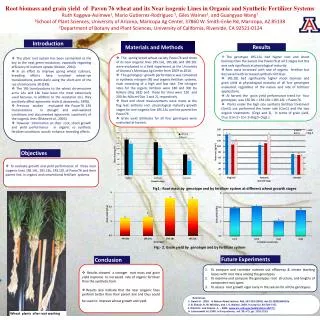
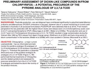
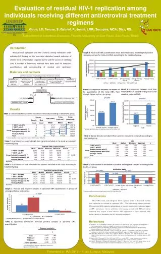
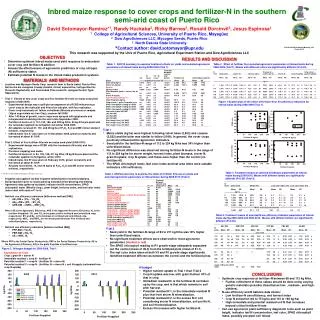
Download
1 / 10
110 likes | 242 Vues
Methods and Materials: Microscopic & Drug Distribution Studies. Presented By: Rich Dominiak Laura Kuczynski John Roszko. February 14, 2006. Microscopic Study Procedure. 1 st – 10 µm thin sections were obtained using a cryogenic microtome set at -25 °C with a microtome knife

E N D
Methods and Materials: Microscopic & Drug Distribution Studies Presented By: Rich Dominiak Laura Kuczynski John Roszko February 14, 2006
Microscopic Study Procedure • 1st – 10 µm thin sections were obtained using a cryogenic microtome set at -25 °C with a microtome knife • 2nd – 1 mL of embedding medium was poured onto the chucks • 30 seconds later it became an opaque solid
Procedure cont’d • Next, about 3 mm x 1 mm x 1 mm of release matrix was taken from various location in the original 1 cm x 1 cm x 1 mm slab. • These pieces of the matrix were then placed on the planed embedding medium, which was in contact to the planed surface.
Procedure cont’d • More hardening medium was added to the planned surface. • Sections 10 µm in thickness were cut and stuck to the microtome knife • They were then taken off of the knife with a glass slide at room temperature
Final Matrix • The final sections were 3 mm x 1 mm x 10 µm. • The 1 mm represents the original depth of the matrix.
Observation • They were observed under scanning electron microscopy (SEM). • Dried to enable high vacuum conditions for SEM.
Drying • Drying Procedure • Water to 100% ethanol • 100% ethanol to 100% amyl acetate • Cooled to 4 °C and filled with liquid CO2 • Exhausted the CO2 vapor while adding CO2liquid • Temperature and Pressure increased over 20 minutes to 40 °C and 55 atm, respectively (when amyl acetate was gone)
Drying cont’d • After 20 min it is assumed all amyl acetate is gone because all the CO2 is vapor • Pressure and Temperature went back to ambient conditions • Sample is ready for analysis
Drug Distribution Studies • Separating matrix into four sections • Removed and frozen on dry ice to terminate drug release • Serially cut with cryomicrotome into four sections for analysis • The samples were set in 0.9% NaCl solution for 3 days • The medium was then filtered and protein concentration was determined by UV spectroscopy at 220 nm