Download
1 / 40
420 likes | 727 Vues
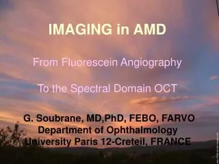
IMAGING in AMD From Fluorescein Angiography To the Spectral Domain OCT. G. Soubrane, MD,PhD, FEBO, FARVO Department of Ophthalmology University Paris 12-Creteil, FRANCE. D iagnosis of CNV: G old standard Leakage through abnormal endothelium and CNV

E N D
IMAGING in AMD From Fluorescein Angiography To the Spectral Domain OCT G. Soubrane, MD,PhD, FEBO, FARVO Department of Ophthalmology University Paris 12-Creteil, FRANCE
Diagnosis of CNV: Gold standard Leakage through abnormal endothelium and CNV Staining of additional tissue or of RPE Evaluation of inner (vessels) and outer (RPE) retinal barriers Diffusible molecule through choriocapillaris Basis for staging of the disease Age Related Maculopathy vs Age related Macular Degeneration Geographic Atrophy vs Choroidal New Vessels FLUORESCEIN ANGIOGRAPHY
Soft drusen Hard drusen Soft drusen Choroidal New Vessels Geographic Atrophy
Choroidal New Vessels • LES 2 TYPES DE C Pre epithelial classic Sub epithelial occult
FA CLASSIFICATION • Age Related Maculopathy (ARM) • RPE changes : hyper or hypopigmentation • Drusen : hard, soft, calcified, reticular pseudo-drusen • Age related Macular Degeneration (AMD) • Atrophy • Choroidal new vessels (CNV) : • classic , • occult (MPS type II), • PED serous, fibrovascular ( MPS type I)
Normal choroidal circulation ARM : Distinction of various material AMD : Dynamic visualisation of Abnormal network and new-vessels Conversion of occult CNV into sub-epithelial CNV Visualization of CNV inside a PED Diagnosis of polypoidal vasculopathy and chorioretinal anastomosis INDOCYANIN GREEN ANGIOGRAPHY
Hard drusen Soft drusen
Sub-epithelial occult CNV • Progressing sub-epithelial occult CNV: • AF : discreteabnormalities without diffusion • ICG : early filling of the central feeder vessel perfusing a neovascular network • within a dark area • late staining of a persistent central plaque
Sub-epithelial occultCNV Natural history:Clinical development of PED
Fibro vascular PED PED Occult
Natural history Sub-epithelial CNV Proliferation of pre-epithelial classic new vessels In 42%, in 2 to 3 years Angiographies : irreplaceable tools • AF : visualization of pre-epithelial classic new vessels • ICG : identification of sub-epithelial occult new vessels
Chorioretinal anastomosis • Severe form of neovascularization • Frequency 15% of AMD (4.5% for Japanese*) 30% of vascularized PED Prognosis for second eye * Am J Ophthalmol. 2007
Idiopathic polypoidal vasculopathy Biomicroscopy
ICG CLASSIFICATION • Age Related Maculopathy (ARM) • RPE changes : hyper or hypopigmentation • Drusen : hard, soft, (reticular pseudo drusen) • Age related Macular Degeneration (AMD) • Atrophy • Choroidal new vessels (CNV) : • Sub epithelial occult with RPE elevation • Vascularized PED • Ingrowth of classic pre epithelial • Fibrovascular PED • CRA, Polyps
Direct and indirect symptoms Accumulation of fluid in all retinal layers Changes in the neurosensory retina especially of the photoreceptors Irregularity or elevation of the RPE Quantification of the abnormalities : retinal thickening or thinning OPTICAL COHERENCE TOMOGRAPHY
CIRRUS SD-OCT SPECTRALIS SD-OCT TOPCON SD-OCT OCT 3 OCT 1 HR-OCT
Outer nuclear layer External limiting membrane Inner segment Interface Outer segment RPE Bruch’s membrane Spectral OCT • Analysis of the outer hyper-reflective layers • - external limiting membrane • - interface OS/IS • - RPE • - Bruchmembrane
Sub- epithelial occult CNV Network
Sub- epithelial occult CNV Fovea Junction OS/IS Irregular, fragmented RPE
Sub- epithelial occult CNV VA 20/50 P3
Sub- epithelial occult CNV Fovea SRF RPE RPE detachment organized with discrete shadowing SRF extensive RPE thinnedand irregular
Sub- epithelial occult CNV Fovea 6 mm RPE detachment • - Elevated RPE • with moderate reflectivity • no marked shadowing • Limited subfoveal SRF • small increase in retinal thickness • foveal flattening
Serous PED b PED with Spectral Domain OCT dépression fovéale Fovea RD DSR DEP PED Occult Fibro vascular PED
CRA Large Cysts CRA with Spectral OCT FA Late ICG VA: 20/50
Fluid or not fluid • Early detection of fluid • Quantification of retinal thickness • Response to treatment • Outer retinal layers • Visibility of Bruch’s membrane • IS-OS changes • Retinal atrophy • Beginning of choroidal analysis • In the future Adaptive Optics • PED OCT CLASSIFICATION
Evolution of imaging for neovascular AMD OCT ICG Angiography SD-OCT Huang Puliafito Gass Flower Yannuzzi Drexler Coscas Fluorescein Angiography 1967 2000 2006 - 2009 Classic, Occult CNV, FV PED Subepithelial CNV
Submacular surgery Laser photocoagulation PDT with verteporfin Anti-VEGF therapy SST MARINA ANCHOR Creteil MPS reports TAP report VIP reports VIM VISION PIER Evolution of therapy for neovascular AMD 1980 2000 2006 - 2009 Improve vision Halt progression PDT = photodynamic therapy