Download
1 / 19
230 likes | 715 Vues
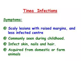
Tinea Infections Symptoms: @ Scaly lesions with raised margins, and less infected centre @ Commonly seen during childhood. @ Infect skin, nails and hair. @ Acquired from domestic or farm animals. Aetiology: @ Caused by filamentous fungi which

E N D
Tinea Infections Symptoms: @ Scaly lesions with raised margins, and less infected centre @ Commonly seen during childhood. @ Infect skin, nails and hair. @ Acquired from domestic or farm animals
Aetiology: @ Caused by filamentous fungi which invade the stratum corneum. @ Three genera are responsible: * Trichophyton, * Microsporum * Epidermophyton (one species: E. floccosum).
Classification: @ Tinea pedis (feet): * infects the interdigital spaces (known as 'athlete's foot') * Causes itching, and blisters. * Caused by T. rubrum and T. interdigitale @ Tinea cruris (groin): * Causes genitourinary infection
@ Tinea corporis (body): * Referred to as 'ringworm’. * Infect the arms, trunk and legs. * Caused by T. rubrum @ Tinea capitis (head): * Infects the scalp and hair. * Causes scaling of scalp, severe dandruff, and broken hair. * Rare in adults.
Lab. Diagnosis of Tinea: @ Presence of scales is diagnostic of 'ringworm' @ Wood’s ultraviolet illumination of scalp * Microsporum infections green light. * Not applicable for other genera. @ Specimens: * Skin scales, nail clippings, and hair. * Swabs are unsuitable. * Skin scales are obtained from edge not the centre of the lesion
@ Microscopy: Specimens are digested in 10-20% potassium hydroxide. @ Culture: Specimens are cultured on Sabouraud agar containing antibiotics and cycloheximide to suppress other organisms. Incubation at room temp. may take up to 3 weeks.
Other Skin Infestations presenting with a rash or itching: @ Superficial candidiasis: * Infects skin, nail or feet @ Pityriasis versicolor: * Caused by Malassezia furfur (yeast) * Shows by hypo-or hyperpigmented macules. * It is not itchy. * Diagnosed clinically & by microscopy
@ Scabies: * Caused by insect Sarcoptes scabiei. * Affects finger clefts, forearms and genital area. * Acquired by sexual close contact. @ Pediculosis pubis: * Caused by crab lice * Affects the head, body, pubic area. @ Viruses: * Herpes, enteroviruses, Measles, Rubella
Classifications of dermatophytes According to Source