Download
1 / 142
1.76k likes | 2.86k Vues
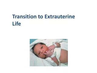
Transition to Extrauterine Life. Pulmonary System Transition. Function of respiration switches from the placenta to the lungs Factors responsible for onset of breathing Hypercapnia Hypoxia Acidosis Environment (cold, light, noise) Fluid in the lungs must be cleared

E N D
Pulmonary System Transition • Function of respiration switches from the placenta to the lungs • Factors responsible for onset of breathing • Hypercapnia • Hypoxia • Acidosis • Environment (cold, light, noise) • Fluid in the lungs must be cleared • Pulmonary arterioles dilate, PVR level falls
Respiratory Adapations • Mechanical changes • Chemical changes • Thermal changes • Sensory changes
Cardiac System Transition • Pressure in right side of the heart falls and pulmonary venous return to left atrium increases • Foramen ovale closes due to these changes • Ductus arteriosis constricts and closes functionally by 96 hours • Ductus venosis constricts and closes functionally by two to three days
Cardiovascular Adaptations • Decreased pulmonary vascular resistance and increased blood flow • Increased systemic pressure and closure of ductus venosus • Increased left atrium and decreased right atrium pressure • Closure of foramen ovale • Reversal of blood flow through ductus arteriosus and increased PO2 • Closure of ductus arteriosus
Figure 28–4 Transitional circulation: conversion from fetal to neonatal circulation.
Figure 28–6 Fetal-neonatal circulation. A, Pattern of blood flow and oxygenation in fetal circulation. B, Pattern of blood flow and oxygenation in transitional circulation of the newborn. C, Pattern of blood flow and oxygenation in neonatal circulation.
Figure 28–6 (continued) Fetal-neonatal circulation. A, Pattern of blood flow and oxygenation in fetal circulation. B, Pattern of blood flow and oxygenation in transitional circulation of the newborn. C, Pattern of blood flow and oxygenation in neonatal circulation.
Figure 28–6 (continued) Fetal-neonatal circulation. A, Pattern of blood flow and oxygenation in fetal circulation. B, Pattern of blood flow and oxygenation in transitional circulation of the newborn. C, Pattern of blood flow and oxygenation in neonatal circulation.
Fetal Laboratory Value Changes • Decreased erythropoietin production • Rise of hemoglobin concentration • Physiologic anemia of infancy • Leukocytosis • Decreased percentage of neutrophils
Thermoregulation • Body heat lost easily due to large body surface area in relation to weight • Limited neonatal fat stores • Limited capacity for heat production • Brown-fat metabolism is primary heat source
Thermoregulation (continued) • Normal axillary temperature is 97°F–99.5°F • Hypothermia is <97.0 • Goal is to keep infant in a neutral thermal environment
Thermogenesis in the Newborn • Large body surface area compared to mass • Types of heat loss • Convection • Radiation • Evaporation • Conduction
Figure 28–9 Methods of heat loss. A, Convection. B, Radiation. C, Evaporation. D, Conduction.
Figure 28–9 (continued) Methods of heat loss. A, Convection. B, Radiation. C, Evaporation. D, Conduction.
Figure 28–9 (continued) Methods of heat loss. A, Convection. B, Radiation. C, Evaporation. D, Conduction.
Figure 28–9 (continued) Methods of heat loss. A, Convection. B, Radiation. C, Evaporation. D, Conduction.
Types of Bilirubin • Unconjugated bilirubin • Conjugated bilirubin • Total bilirubin
Conjugation and Excretion of Bilirubin • Bilirubin is transported in blood via albumin • Bilirubin is transferred into the hepatocytes • Attachment of unconjugated bilirubin to glucuronic acid • Excreted into bile ducts, then into the common duct and duodenum • Bacteria transform it into urobilinogen and stercobilinogen • Bilirubin is excreted in urine and stool
Physiologic Jaundice • Accelerated destruction of fetal RBCs • Increased amounts of bilirubin delivered to liver • Inadequate hepatic circulation • Impaired conjugation of bilirubin • Defective uptake of bilirubin from the plasma • Defective conjugation of the bilirubin
Physiologic Jaundice (continued) • Increased bilirubin reabsorption • Defect in bilirubin excretion • Increased reabsorption of bilirubin from the intestine
Liver Adaptations • Iron content stored in liver • Low carbohydrate reserves • Main source of energy is glucose • Liver begins to conjugate bilirubin • Lack of intestinal flora results in low levels of vitamin K
GI Adaptations • Sufficient enzymes except for amylase • Digests and absorbs fats less efficiently • Salivary glands are immature • Stomach has capacity of 50-60 mL • Cardiac sphincter is immature
Fluid and Electrolyte Balance • Less able to concentrate urine • Limited tubular reabsorption of water • Limited excretion of solutes • Limited dilutional capabilities
Immunologic Responses in the Newborn • IgG – passive acquired immunity via placenta • IgM – usually not passively transferred • Elevated levels may indicate fetal antigenic activity in utero • IgA – passive acquired immunity via colostrum
Periods of Reactivity • First period of reactivity • Sleep phase • Second period of reactivity
Behavioral and Sensory Capabilities • Habituation • Orientation • Auditory • Olfactory • Tasting and Sucking • Tactile
Dry infant, remove wet blankets Apply a hat and warm blankets Avoid placing infant on cold surfaces Avoid placing infants in drafts Nursing Interventions to Prevent Hypothermia
Nursing Interventions to Prevent Hypothermia (continued) • Use heat source when bathing infants • Place under radiant warmer if temperature is unstable
Metabolic Transition • Infant’s source of nutrition from the placenta terminates at birth • Blood sugar reaches its lowest point one to three hours after birth • Glucose stabilizes by four to six hours after birth • Range of 45–80 mg/dl is normal
At birth abdomen is flat and bowel sounds are absent Abdomen becomes rounded and soft with onset of respirations Bowel sounds usually audible within 15 minutes of birth Gastrointestinal System
First minutes after birth Characteristics Alert, active, sucking activity, tachycardia, tachypnea, transient rales and nasal flaring Implications for the family Infant alert and responsive Allow quiet time for family to be together Introduce breastfeeding First Period of Reactivity
Follows first period of reactivity Characteristics Less alert and active, sleep may occur, vital signs normalize Period of Decreased Activity
Period of Decreased Activity (continued) • Implications for the family • Family may stay together or infant may be taken to nursery for assessment • Opportunity for parents to have quiet time • Mother may use this time to rest
Second Period of Reactivity • Infant awakens and shows increased responsiveness to the environment • Characteristics • Peristalsis increases and meconium may be passed, gagging, spitting up • Implications for the family • Allow time together if mother is rested • Parents may begin to have questions or need assistance with newborn care
Asphyxia • Arises from inadequate or absent respiration • Impairment of oxygen/carbon dioxide exchange • Hypoxemia, hypercarbia, respiratory acidosis • Assessment findings • Poor tone, gasping or absent respirations, bradycardia, cyanosis, low Apgar score
Asphyxia (continued) • Management • Tactile stimulation • Positive pressure ventilation with 100% oxygen
Meconium Staining • Caused by distress, usually asphyxia • Risk is that fetus/infant may aspirate • Obstruction, chemical pneumonia may result • Assessment findings • Respiratory distress, hypoxemia • Prevention • Suctioning nose/mouth before delivery of the chest • Appropriate suctioning post delivery
Transient Tachypneaof the Newborn • Characteristics • Grunting, retracting, tachypnea • Risk factors • Cesarean delivery, precipitous delivery • Management • Oxygen therapy • IV fluids • Short-term ventilation • Antibiotics if sepsis is suspected
Hypoglycemia • Plasma glucose level below 40 mg/dl • Assessment findings • Jitteriness, tremors, apnea, cyanosis, lethargy • Risk factors • SGA, preterm, perinatal stress, IDM, sepsis
Hypoglycemia (continued) • Management • Early feeding of infants at risk • Keep infant warm • Glucose by nipple, gavage, or IV • Recheck blood glucose 30 minutes after feeding
Transition of the Premature Infant • Pulmonary system • Inadequate alveolar development, lack of surfactant • May require ventilatory support • Administration of surfactant • Cardiac system • Persistent ductus arteriosis (PDA) • Indomethacin given to facilitate closure
Resuscitation and Stabilization in the Delivery Room • Dry and provide warmth, tactile stimulation • Clear airway • Resuscitation for compromised infants • Place under radiant warmer, stimulate • Position to ensure a patent airway • Suction using appropriate technique • Evaluate respirations, heart rate, color