Download
1 / 76
760 likes | 942 Vues
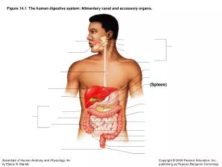
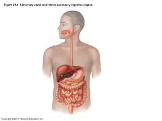
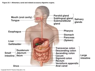
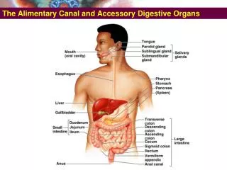
The Alimentary Canal and Accessory Digestive Organs. Abdominal Quadrants. A simpler scheme defining four quadrants. Nine Regions of Anterior Abdominal Surface. Nine Regions of Anterior Abdominal Surface. How regions relate to abdominal viscera.

E N D
Abdominal Quadrants • A simpler scheme defining four quadrants
Nine Regions of Anterior Abdominal Surface • How regions relate to abdominal viscera
Location of Organs in the Abdominopelvic Regions • Hypogastric- large intestine, sm intestine, bladder • Umbilical- sm and lg intestine • Epigastric- stomach, liver, spleen, pancreas • Right hypochondriac- liver • Left hypochondriac- stomach, liver, spleen, pancreas • Right lumbar- large and small intestine • Left lumbar- large and small intestine • Right iliac- large intestine, cecum • Left iliac- large intestine
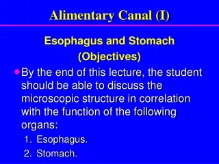
The Small Intestine • The Intestinal Wall • Mucosa has transverse folds, plicaecirculares. • Plicae have small projections, villi. • Both increase surface area of mucosa for absorption. • Each villus has a lymphatic capillary, a lacteal.
The Small Intestine • Intestinal Secretions • Intestinal glands secrete • Intestinal juice • Moistens chyme • Buffers stomach acid • Dissolves digestive enzymes • Dissolves products of digestion • Mucus • Hormones
The Small Intestine • Intestinal Hormones • Gastrin • Secretin • Cholecystokinin (CCK) • Gastric Inhibitory Peptide (GIP)
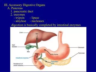
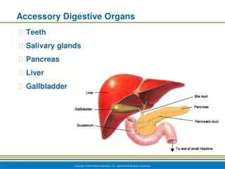
The Small Intestine • Digestion in the Small Intestine • Most enzymatic digestion and absorption occurs in the small intestine • Digestive enzymes and buffers are released by: • Pancreas • Liver • Gall bladder
The Small Intestine – Gross Anatomy • Longest portion of the alimentary canal • Site of most enzymatic digestion and absorption • Three subdivisions • Duodenum • Jejunum • Ileum
The Small Intestine – Gross Anatomy • Plays Important role in digestion and absorption • Mucosa of SI produce few enzymes, and buffer to neutralize chime • Divided into three part: • Duodenum – receives chime from stomach, bile from gall bladder, and digestive secretion from pancreas • Digestion continues in duodenum • Jejunum –ileum: digestion and absorption takes place here • Ileocecal sphincter • Transition between small and large intestine
Sectional Differences of Small Bowel • Autopsy: 7M • Life: 4.5~5M • Duodenum • Shortest, widest, 25cm, c-shape • Jejunum • 2/5, coiled spring, feathery • Ileum • 3/5, smoother
The Duodenum • Receives digestive enzymes and bile. • Main pancreatic duct and common bile duct enter duodenum. • Sphincters control entry of bile and pancreatic juices.
The Duodenum of the small intestine • The first • The shortest • The widest • The most deeply lying, • The least accessible, • Anterior surface is covered in peritoneum. • Posterior surface is not covered in peritoneum.
The Duodenum • Divided into four parts: • Superior- the first • Descending- the second • Horizontal- the third • Ascending- the fourth • Duodenal flexure
The Duodenum • Divided into four parts: • Superior- the first • Descending- the second • Horizontal- the third • Ascending- the fourth • Duodenal flexure
The Duodenum • Superior part: • Duodenal ampulla or cap • Pylorus protrudes in it. • Forms the inferior margin of the epiploic foramen. • Lesser omentum attaches to its upper margin. • Greater omentum attaches to its lower margin.
The Duodenum Relations of superior part • Anteriorly • Quadrate lobe of liver • Gallbladder • Posteriorly • Common bile duct • Gastroduodenal a. • Hepatic portal v. • Inferior vena cava • Superiorly • Omental foramen • Inferiorly • Head of pancreas
The Duodenum • Descendingpart: • Theanteriorsurface is coveredwithperitoneum. • Exceptalongtheattachmentline of thetransversemesocolon. • Thehead of thepancreas is in directcontactwith it. • Thecommon bile ductandthemainpancreaticductopenintoitslumen. • Majorduodenalpapillaandminorduodenalpapillaand
The Duodenum • Descendingpart: • Theanteriorsurface is coveredwithperitoneum. • Exceptalongtheattachmentline of thetransversemesocolon. • Thehead of thepancreas is in directcontactwith it. • Thecommon bile ductandthemainpancreaticductopenintoitslumen. • Majorduodenalpapillaandminorduodenalpapillaand
The Duodenum Relations of descending part • Anteriorly • Liver • Transverse colon and transverse mesocolon • Loops of small intestine • Posteriorly • Right renal hilum and ureter • Right renal vessels • Medially • Head of pancreas • Common bile duct and pancreatic duct • Laterally • Right colic flexure
The Duodenum • Horizontalpart: • Crossestotheleft in front of. • L3 vertebra • Theinferior vena cava • The aorta • Continueswiththeascendingpart in front of the aorta. • Theanteriorsurface is coveredwithperitoneum. • Exceptalongtheattachmentline of themesentery.
The Duodenum • Horizontalpart: • Crossestotheleft in front of. • L3 vertebra • Theinferior vena cava • The aorta • Continueswiththeascendingpart in front of the aorta. • Theanteriorsurface is coveredwithperitoneum. • Exceptalongtheattachmentline of themesentery.
The Duodenum Relations of horizontal part • Superiorly • Head of pancreas • Inferiorly • Loops of small intestine • Anteriorly • Radix of mesentery • Superior mesenteric a. and v. • Posteriorly • Right ureter • Inferior vena cava • Abdominal aorta
The Duodenum Relations of horizontal part • Thesuperiormesenteric a. / v. • Enters / leavestheroot of themesentery. • Bothvessels: • Crossthehorizontalsegmentanteriorly. • Thesevesselsmaycompresstheduodenum, leadingtodistention of theproximalduodenumandstomach. • Known as SMA syndrome, manifestbyabdominalpain, nausea, andvomiting.
The Duodenum • Ascendingpart: • ReturnstoL2 vertebra. • Runs on theleftside of the aorta. • Forms sharp duodenojejunal flexure • Theanteriorsurface is coveredwithperitoneum. • Exceptalongtheattachmentline of themesentery.
The Duodenum Relations of ascending part • Right — Head of pancreas and abdominal aorta • Left — left kidney and ureter
The Duodenum • Suspensorymuscle of theduodenum: • Securestheduodenumtotheposteriorabdominalwalland has twoparts: • Onederivedfromthediaphragm, whichcontainsstriatedmuscle. • Theotherpartderivedfromtheduodenalwall, whichcontainssmoothmuscle.
The Jejunum and Ileum • Theyarespecialisedfor • Absorption of digestedfood, • Vitamins, • Electrolytes • Theyfill • Infracoliccompartment • Pelviccavity • Peritoneum • Completelyinveststhem • Exceptalong a narrowstripwhere it becomescontinuouswiththetwolayers of peritoneum, themesentery.
The Jejunum and Ileum • Jejenum: • Thejejenum is slightlywiderthantheileum. • Themucosa is thrownintocircularfolds in thejejenum, whereascircularfolds in theileumaresmallandsparse. • Ileum has distinctivelymphoidfollicles (Peyer’spatches); suchlargefolliclesareabsent in thejejenum.
The Jejunum and Ileum • Jejunum begins at duodenojejunalflexure, no clear demarcation to ileum. • Jejunum has long vas recta, large plicae, thick walls, transparent mesentery. • Ileum has short vasa, small plicae, thin walls, fat in the mesentery.
The Large Intestine (Intestinum Crassum) • Starts at the ileocecal junction. • Ends at the anus. • Approximately 1.5m long. • Consists of • Cecum • Colon • Rectum • Anal canal
The Large Intestine • Overview of the Large Intestine • Reabsorbs water and compacts feces • Absorbs vitamins made by bacteria • Stores feces before defecation • Consists of three parts • Cecum:Attaches to vermiform appendix • Colon • Rectum:Terminates in anal canal
The Large Intestine • Mechanical digestion • Haustral churning • Peristalsis • Mass peristalsis – drives contents of colon toward rectum • Chemical digestion • Final stage of digestion through bacterial action • Ferment carbohydrates, produce some B vitamins and vitamin K • Mucus but no enzymes secreted • Remaining water absorbed along with ions and some vitamins
The Large Intestine • Functions of the Large Intestine • Absorption • Water • Ions • Vitamins • Organic wastes • Bile salts • Toxins • Bacterial growth
Gross Anatomy of Large Intestine • Subdivided into • Cecum, vermiform appendix, colon, rectum, anal canal • Special features of large intestine • Teniae coli • Thickening of longitudinal muscularis • Haustra • Puckering created by teniae coli • Epiploic appendages • Fat-filled pouches of visceral peritoneum
Gross Anatomy of Large Intestine • Cecum • Blind pouch • Beginning of large intestine • Vermiform appendix • Contains lymphoid tissue • Neutralizes pathogens • Colon • Divided into distinct segments • Ascending, transverse, descending, and sigmoid colon
Gross Anatomy of Large Intestine • Rectum • Descends along the inferior half of the sacrum • Anal Canal • The last subdivision of the large intestine • Lined with stratified squamous epithelium
The Large Intestine (Intestinum Crassum) • Converts the liquid contents of the ileum into semisolid feces by the time the sigmoid colon is reached. • This is accomplished by the absorption of fluid and electrolytes. • The transverse and sigmoid colon possess considerable mobility. • The ascending, descending colon, and rectum are fixed to the posterior wall of abdominal cavity. • The cecum and appendix are completely peritoneal.
The Large Intestine (Intestinum Crassum) • The longitudinal muscle of the cecum and colon is gathered into three narrow ribbonlike bands called teniae coli. • Omental • Free (anterior) • Mesenteric • Teniae are shorter than the gut tube itself. • This causes series of sacculations called haustra coli. • Appendices epiploicae • Bodies of fat enclosed by peritoneum, hanging from the teniae.
Gross Anatomy of Large Intestine • Caecum • Dilated portion • Blind end inferiorly • Continuous superiorly as the ascending colon • The terminal ileum opens into the large intestine Just below the junction of the caecumand ascending colon • The opening is guarded by ileocaecal valve. • The blind end has appendix attached to it .
Gross Anatomy of Large Intestine • Caecum • Dilated portion • Blind end inferiorly • Continuous superiorly as the ascending colon • The terminal ileum opens into the large intestine just below the junction of the caecumand ascending colon • The opening is guarded by ileocaecal valve. • The blind end has appendix attached to it .
Ileocecal region: • Different positions of the appendix:- • Retrocecal (the commonest). • Subcecal. • Anteileal. • Retroileal. • Pelvic.
Gross Anatomy of Large Intestine • Caecum • Locatedjustabovethelateralthird of theinguinalligament. • Can be palpated in therightiliac fossa. • Itsliquidcontents do not offerresistancetothepalpatingfingers. • Consequently, therightiliac fossa feelsemptytopalpation. • Ileocecalvalve: not veryefficient, backflowthrough it intotheileum can occurwhenthecolonandcecumarefilled.
Gross Anatomy of Large Intestine • Ascending colon • Passes upwards from caecum to the level of the liver – turns to the left –hepatic flexure – becomes • Transverse colon • A loop curved downwards across the • abdomen upto spleen – turns • downwards – splenic flexure– • becomes • Descending colon • Passes down from splenic flexure • situated on the left side of the • abdomen. • In the true pelvic cavity it is called • sigmoid or pelvic colon