Endocrinology:
Endocrinology:. Chapter 11. Endocrine System Function. Major control and communication system Controls… body fluid composition and volume nutrient levels growth and development reproduction physiological cycles (“biological clocks”). Nervous vs. Endocrine Control.

Endocrinology:
E N D
Presentation Transcript
Endocrinology: Chapter 11
Endocrine System Function • Major control and communication system • Controls… • body fluid composition and volume • nutrient levels • growth and development • reproduction • physiological cycles (“biological clocks”)
Nervous vs. Endocrine Control • The nervous system controls rapid, precise responses (ex. reflex) • The endocrine system controls activities that require long duration (ex. body growth) • energetically more efficient • Specific actions of chemical messengers are at the level of the target cell • These two systems interact and regulate each other
The Endocrine System • Endocrine glands • Lack ducts • Secrete products into the interstitial fluid • Endocrine organs may be solely endocrine or multifunctional
Major Endocrine Organs • Pituitary and Hypothalamus • Adrenal glands (2) • Thyroid • Parathyroid glands (4) • Pancreas • Ovaries, Testes Fig 11.1
Additional Endocrine Organs Table 11.1
Hormones Chemicals that are broadcast throughout the body which induce physiological changes in specific target cells.
Hormone Classes • Amines • hormones derived from tyrosine and tryptophan • adrenal medulla hormones, thyroid hormones, pineal gland hormones • Peptide Hormones • made from polypeptide chains • most hormones (insulin, FSH) • Steroids • derivatives of cholesterol • adrenal cortex hormones, gonadal hormones Figs 11.2, 3.25, 11.3
Mechanism of Action:Steroids & Thyroid Hormones • nonpolar • pass directly through the cell membrane • bind to protein receptor in cytoplasm or in nucleus • protein binds to gene on DNA in the nucleus • stimulates expression of that gene (protein production) Figs 11.4 and 11.6
Mechanism of Action: Peptides and Most Amines • Polar • cannot pass through hydrophobic lipid bilayer • bind to receptor proteins on cell surface • activation of membrane-bound enzymes • production of a second messenger inside the cell • e.g. cAMP • 2nd messenger activates or deactivates various enzymes Figs. 11.8, 11.9 and 11.11
Hormonal Regulatory Mechanisms • Regulating hormone levels • e.g. Negative feedback • Change causes change in opposite direction • e.g. thyroxine/TSH • Regulating tissue response • e.g. down regulation • Decrease # of receptors on target cell with chronically elevated hormone levels Fig 11.16
Hypothalamus-Pituitary Axis • Hypothalamus • part of the diencephalon • controls release of pituitary hormones • Neural control of endocrine function • Pituitary gland • extends from the inferior surface of the hypothalamus • Two distinctive lobes (posterior and anterior) • Linked to hypothalamus by infidiubulum Fig 11.12
Posterior Pituitary • Composed of nervous tissue • Neurosecretory cells produce two peptide hormones • Released when neurons undergo an AP Fig 11.13
Posterior Pituitary Hormones • ADH (Anti-Diuretic Hormone) • increases reabsorption of H2O by kidneys • induces vasoconstriction in arterioles - BP • stim. by H2O deficit, BP • Oxytocin • Uterine contraction during childbirth • milk letdown during breast feeding • male function unclear ( occurs during ejaculation) Fig 11.13
Anterior Pituitary • Composed of epithelial cells • Different cell types secrete one of six peptide hormones Fig 11.12
Anterior Pituitary Hormones • TSH (Thyroid Stimulating Hormone) • Synthesis/ release of thyroid hormones • Thyroid growth • ACTH (Adrenocorticotrophin) • Activates adrenal cortex to release glucocorticoids Fig 11.14
Anterior Pituitary Hormones • GH (Growth Hormone, or Somatotropin) • Stimulates secretion of growth factors from various tissues • GF’s timulate growth, protein synthesis, fat breakdown and blood glucose levels • PRL (Prolactin) • breast development and milk production during pregnancy • Modulatory roles in male reproduction and ion balance Figs 11.14, 19.18
Anterior Pituitary Hormones • LH (Luteinizing Hormone) • Females • ovulation, regulation of female sex hormones • induces corpus luteum formation after ovulation • Males • regulation of male sex hormones (androgens) Fig 11.14
Anterior Pituitary Hormones • FSH (Follicle Stimulating Hormone) • Females • regulates female sex hormones, egg development • Males • Induces local mediator secretion from Sertoli cells that trigger sperm development Fig 11.14
Hypothalamal Control of the Anterior Pituitary • Hypothalamal neurons produce releasing/inhibiting hormones • Stimulate or inhibit secretion of hormones from the anterior pituitary • Released into Hypothalamo-hypophyseal portal blood vessels Fig 11.15
Hypothalamal Regulatory Hormones • TRH (thyrotrophin-releasing hormone) • stimulates TSH release • CRH (corticotrophin-releasing hormone) • stimulates ACTH release • GHRH (growth hormone releasing hormone) • stimulates GH release • Somatostasin • inhibits GH release • PIH (prolactin inhibiting hormone) • inhibits prolactin release • GnRH (gonadotrophin-releasing hormone) • simulates FSH and LH release
Adrenal Gland • Located above each kidney • Releases hormones in response to stress • Medulla hormones (amines) • Epinephrine & Norepinephrine- similar to effects of sympathetic NS (“flight or fight”) Fig 11.18
Adrenal Gland • Cortex hormones (steroids) • glucocorticoids (blood glucose) • Cortisol – elevates blood glucose and fatty acid levels, inflammation suppression • mineralocorticoids (salt) • Aldosterone – increases K+ secretion and Na+ uptake by kidneys • androgens • (DHEA) – secondary sex character development, sexual behavior Fig 11.18
Glucocorticoid Regulation • Cortisol - helps body cope with stress • Hypothalamus releases CRH • Stimulates ACTH from anterior pituitary • Stimulates cortisol release from adrenal gland • Cortisol inhibits CRH release and desensitizes ant. pit. to its effects Fig 11.20
Thyroid Gland • Produces two groups of hormones • Thyroid hormones (amines) • Thyroxine (T4) and triiodothyronine (T3) - Increase metabolic rate and body heat production • Calcitonin (peptide) • increases bone matrix formation and Ca2+ secretion from kidneys • reduces blood Ca2+ levels Fig 11.21
Thyroxine Regulation • Secretion regulated by the hypothalamus-pituitary axis • Hypothalamus releases TRH • TRH stimulates ant. pituitary to release TSH • TSH stimulates thyroid to secrete T4 • negative feedback of T4 onto ant. pituitary • T4, TSH release Fig 11.16
Thyroid Abnormalities:Hyperthyroidism • Grave’s Disease • production of thyroid stimulating immunoglobin • Mimics TSH function, not subject to negative feedback regulation • Overproduction of thyroid hormones • Symptoms • BMR = body weight, body temp • Hyperexcitability of nervous system • Restless behavior, HR, etc. • Exopthalmos - bulging eyes Fig 11.26
Thyroid Abnormalities:Hypothyroidism • General symptoms • Decreased BMR • Body temperature • Weight gain • Alterness (impaired CNS function) • Easily fatigued
Thyroid Abnormalities:Hypothyroidism • Cretinism • low TH production during infancy • Reduced growth rate (dwarfism) • Severe mental retardation Fig 11.27
Thyroid Abnormalities:Hypothyroidism • Goiter Formation • Reduced thyroid hormone production due to iodine deficiency • TSH production • Abnormal thyroid growth Figs 11.24, 11.25
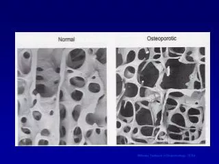
Parathyroid Glands • Four small organs located on posterior surface of the thyroid • Secrete parathyroid hormone (PTH) • promotes bone matrix breakdown • Reduces Ca2+ secretion in the kidneys • Elevates Ca2+ in blood Figs 11.28, 11.29
Pancreas • Both an endocrine organ and digestive organ • Endocrine cells located in Islets of Langerhans • Contain 2 cell types • cells - secrete glucagon • cells - secrete insulin • Important in regulating glucose levels of the blood Fig 11.30
Insulin • Induces glucose uptake and utilization by cells (esp. muscle and liver) • Lowers blood glucose levels • promotes removal of glucose from blood • Promotes formation of glycogen • polymer of glucose for storage • Promotes conversion of glucose into fat in adipose tissue • Stimulates amino acid uptake by cells and protein formation
Insulin Regulation • Blood glucose level is the major factor controlling insulin and glucagon secretion • glucose → insulin → glucose • glucose → insulin → glucose • Maintenance of blood glucose at homeostatic levels via negative feedback Fig 11.31
Glucagon • Secreted when blood glucose levels are VERY LOW • Increase in blood glucose: • Activates liver enzymes to convert glycogen into glucose • Stimulates breakdown of stored fat and release of fatty acids into blood • used as secondary energy source • Opposes the actions of insulin
Diabetes Mellitus • Insulin deficiency or excessive tolerance • cells do not take up glucose • results in excess glucose in blood (hyperglycemia) • Problems • dehydration • lose excessive water from urination • blood volume/pressure problems • starvation - body cannot use glucose • break down of fats, formation of ketone bodies • metabolic acidosis
Diabetes Mellitus Two types: • Type-I (Insulin-dependent, juvenile-onset) • Degeneration of b-cells • no endogenous insulin • Must give exogenous insulin • Type-II (non-insulin dependent, adult-onset) • Cells desensitized (tolerant) of diabetes • Often due to obesity • controlled by regulating dietary glucose
Hypoglycemia • Overproduction of insulin or hypersensitivity • Reactive hypoglycemia • -cells overproduce insulin in response to increased glucose levels • too much glucose driven into cells from blood • depressed brain function