Download
1 / 32
350 likes | 927 Vues
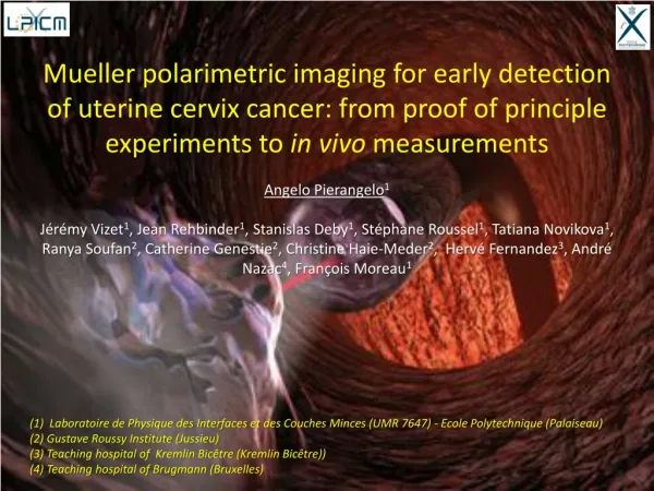
Mueller polarimetric imaging for early detection of uterine cervix cancer: from proof of principle experiments to in vivo measurements Angelo Pierangelo 1

E N D
Mueller polarimetric imaging for early detection of uterine cervix cancer: from proof of principle experiments to in vivo measurements Angelo Pierangelo1 Jérémy Vizet1, Jean Rehbinder1, Stanislas Deby1, Stéphane Roussel1, Tatiana Novikova1, Ranya Soufan2, Catherine Genestie2, Christine Haie-Meder2, Hervé Fernandez3, André Nazac4, François Moreau1 (1) Laboratoire de Physique des Interfaces et des Couches Minces (UMR 7647) -EcolePolytechnique (Palaiseau) (2) Gustave Roussy Institute (Jussieu) (3) Teachinghospital of Kremlin Bicêtre (Kremlin Bicêtre)) (4) Teachinghospital of Brugmann (Bruxelles)
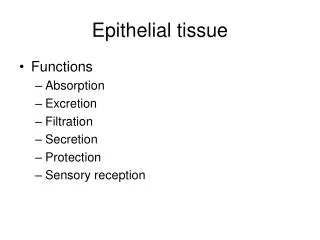
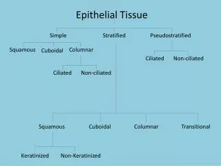
Epithelial cancers Epithelial tissue Epithelium Connective tissue
Epithelial cancers Epithelial cancer development Epithelial tissue Invasive cancer Epithelium Carcinoma in situ Epithelium Connective tissue Cells genetically altered Severe dysplasia Hyperplasia Vessels Connective tissue (collagen, elastin…) Time 90% of cancers
Epithelial cancers Epithelial cancer development Epithelial tissue Invasive cancer Epithelium Carcinoma in situ Epithelium Connective tissue Cells genetically altered Severe dysplasia Hyperplasia Vessels Connective tissue (collagen, elastin…) Time 90% of cancers Detection of precancerous stage Recovery of patients in 95% of cases
Cervical cancer • Major health problem: the second most common female cancer in the world with 275 000 deaths per year, mainly in developing countries (but not only) • Slow evolution (10 - 20 years): ideal case for screening • Direct access to cancer • Model for other epithelial cancers
Cervical cancer screening Current practice Colposcopy Conization Colposcopy : inefficient technique (generating supplementary costs) (60-70% sensitivity and 50% specificity for high-grade dysplasia detection) Visualization of dysplastic areas for biopsy is very difficult and operator dependent Resection margins to achieve a cone biopsy (conization) are poorly defined Significant improvement of colposcopy can make the early detection and surgery both more effective and affordable
Polarization of light Electric field (Polarization) Magnetic field Depolarized light Elliptical polarization Linear polarization y y y x x x
Polarimetric properties Retardance Depolarization Diattenuation Introduction of « disorder » in the evolution of the electric field Phase difference between two polarization eigenstates Difference of transmission for two polarization eigenstates slow fast Birefringence: refraction index varies with polarization Dichroïsm: Absorption coefficient varies with polarization Stochastic process (multiple-scattering) • Biological origins : • Organelles, cytoplasm, fibers… • Biological origins : • Single scattering • Transmission anisotropy of some molecules • Biological origins : • Collagen, elastin, muscle fibers (myosin, actin…)
Mueller polarimetry Stokes vector 4-component vector which describes completely the polarization of light Intensity measurements Mueller matrix M Complete polarimetric response of a sample (including depolarization) Decompositions based on hypothesis on the physical structure of the sample Lu and Chipman [1] : M= MdMRMDReverse : M = MDrMRrMdrSymmetric: M = MD2MR2Mds MR1MD1… [1] : “Interpretation of Mueller matrices based on polar decomposition” , S.-Y. Lu and R. A. Chipman, JOSA A 13 (5)(2012)
Mueller polarimetric imaging Mueller polarimetric imager PSG : 4 probing polarization states Analysis through 4 different configurations of the PSA Probing polarization states Backscattered polarization states 16 measured intensities PSG (Polarization State Generator): linear polarizer, followed by retarders with changing characteristics over time (rotating waveplates, liquid crystal cells, etc...) PSA (Polarization State Analyzer): the same elements as PSG, but in the reverse order Mueller matrix image
Mueller polarimetric imaging …on “ad hoc” samples ex vivo Quartz plate Milk 2cm Tilted glass Mueller Matrix Lu-Chipman decomposition Dichroism Depolarization Retardance
Mueller polarimetric imaging …on biological samplesex vivo Colon (500nm) Uterine cervix (500nm) 5cm 2cm Cancerous zone Pure Depolarizer (diagonal matrix) M22=M33>M44 Rayleigh Scattering Depolarization Retardance Dichroism (non-diagonal matrix)
Mueller polarimetric imaging • Mueller polarimetric imaging : • is sensitive to the microscopic morphology of the tissue • is realized using simple optical elements in the visible range (450 – 700 nm) :different depths in the tissue can be reached using different wavelengths (due to different light absorption by hemoglobin for different wavelengths, hemoglobin being more absorbing for shorter wavelengths); • is well suited for «full field»(few cm²) modality (fundamental for in vivo applications) • can be easily implemented at low cost
Application to cervical cancer 2013 Ex vivo proof of principle measurements 1 Full field Mueller imaging polarimeter ~10 samples analyzed
Application to cervical cancer 2013 2013-2017 Ex vivo proof of principle measurements Ex vivo statistical evaluation In vivo preliminary results 3 Full field Mueller imaging upgraded polarimeters 1 monochromatic Mueller colposcope (550nm) 1 Full field Mueller imaging polarimeter ~10 samples analyzed ~100 samples analyzed ~15 patients analyzed
Application to cervical cancer 2013 2013-2017 2018-2020 Ex vivo proof of principle measurements Ex vivo statistical evaluation In vivo preliminary results In vivo statistical evaluation 3 Full field Mueller imaging upgraded polarimeters 1 monochromatic Mueller colposcope (550nm) Color Mueller colposcope (450 - 550 - 650nm) 1 Full field Mueller imaging polarimeter ~10 samples analyzed ~100 samples analyzed ~15 patients analyzed ~300 patients to analyze
Application to cervical cancer Ex vivo analysis 2cm Characteristics Field of view ≈ 5 x 4 cm² Wavelengths : 400 nm to 700 nm (50 nm step) Time required to acquire a full Mueller matrix Image : several tens of seconds Mueller matrix image and polarimetric parameters
Application to cervical cancer Ex vivo proof of principle measurements Depolarization (550nm) Retardance (550nm) Conventional image CIN 3 2cm Healthy Glandular 1 90° 0° 0 • Healthy zones are characterized by a strong Retardance (~60°) and Depolarization (~1) • Abnormal zones are characterized by a very low Retardance (< 10°) • Different degrees of Depolarization enable to distinguish malignant lesions from benign transformations of the cervix A. Pierangeloet al. Opt.Exp. (2013) J. Rehbinder et al. J. Biomed. Opt. (2015) Prix de l’innovation de l’Ecole Polytechnique 2012
Application to cervical cancer Ex vivo statistical evaluation Statistical evaluation of sensitivity and specificity of the technique using Retardance as a polarimetric diagnostic parameter to distinguish healthy tissue from severe dysplasia (CIN3) on 25 pieces of conizations Automatic reconstruction of the histological mapping Cut A Cut C Cut E 1 2 3 4 1 3 4 5 2 5 Cut B Cut D Histology is the gold standard for the diagnosis of pre-cancerous lesions
Application to cervical cancer Ex vivo statistical evaluation Histological diagnosis Pre-sliced Polarimetric image
Application to cervical cancer Ex vivo statistical evaluation • Uncertainty positioning cut (~1mm) : this introduces an error of 10 – 15% on sensitivity and specificity • We selected subzones between two adjacent lines labeled by the same histological diagnosis
Application to cervical cancer Ex vivo statistical evaluation Comparison of polarimetric imaging with the gold-standard Conventional colposcopy : Sp<50% Less than 50% of healthy zones are properly identified Conventional colposcopy : Se~60-70% 60-70% of cancers detected
Application to cervical cancer Ex vivo statistical evaluation • We fixed a threshold Rs for Retardance • We considered that : • pixels with R>Rshealthy tissue • pixels with R<Rsprecancerous stage • True positive TP are the pixels with R<Rs detected as abnormal zones by pathologists • We calculated Se et Sp by varying Rs
Application to cervical cancer Ex vivo statistical evaluation J. Rehbinder et al. J. Biomed. Opt. (2015)
Application to cervical cancer Microscopy and interpretation of the results CIN3 Healthy The comparison between Mueller polarimetric microscopy and SHG microscopy proved that the strong anisotropy observed in healthy cervix is due to the existence of a highly ordered layer in the connective tissue under the epithelium Strong Retardance of healthy tissues is the signature of collagen Modeling of anisotropic media in progress S. Bancelin et al. Opt. Express 22(19), 22561 (2014)
Application to cervical cancer In vivo measurements Mueller polarimetric colposcope (In vivo) Mueller polarimetric macroscope (Ex vivo) Linear polarizers Analyzed light Incident light 16 images in 20s 16 images in 1.6s
Application to cervical cancer In vivo measurements Intensity image Retardance Depolarization Algebraic methods (LPICM) Adaptative polarimetry (SPIM) Statstical approach (ICUBE, Arizona University) Image obtained combining 550 and 650 nm 0 1 0° 90° Presence of a precancerous lesion (CIN3) confirmed by the biopsy • Healthy zones are characterized by a strong Retardance (~80°) and Depolarization (~0.8) • Abnormal zones are characterized by a very low Retardance (< 10°) • Different degrees of Depolarization enable to distinguish malignant lesions (CIN3) from benign transformations of the cervix (glandular tissue) • These results are compatible with ex vivo In vivomeasurements on a cohort of patients from January 2018 to January 2020 in hospital setting(CHU Kremlin Bicêtre, CHU Brugmann)
Conclusions and perspectives New version of colposcope that is compact, user friendly, fast (16 images in 0.5s) and enables simultaneous acquisition of images at 450, 550 and 650nm December 2017 Measurements by using Mueller colposcopy in vivo on a cohort of patients in two different hospitals (CHU of the Kremlin Bicêtre and CHU Brugmann of Brussels) January 2018 Modeling of anisotropic tissues (Monte Carlo) In progress Exploration of new applications…
New perspectives and applications • Cancerous pathologies • Gastroenterology • Pneumology • Gynecology (Uterus…) • Urology • Dermatology • … • Other non-cancerous pathologies • Preterm delivery (30 patients analyzed at CHU Brugmann of Bruxelles) • …
New perspectives and applications • … towards Mueller polarimetric endoscopy Mueller Microendoscope (collaboration LPICM – Xlim) Retardance (630nm) Microscopy Optical fiber 1mm Scanning Piezoelectric Muscular tissue of colon 0° 120°
New perspectives and applications Challenge: Mueller polarimetric rigid endoscope …in progress Gynecology Mininvasive surgery
Thanks to: Special thanks to: Antonello De Martino