Unveiling Global Form Processing in Macaque Visual Cortex Through Glass Patterns and fMRI Analysis
Explore how Glass patterns activated visual cortex in anesthetized macaque monkeys, revealing areas for global form processing through fMRI imaging with controlled stimuli.

Unveiling Global Form Processing in Macaque Visual Cortex Through Glass Patterns and fMRI Analysis
E N D
Presentation Transcript
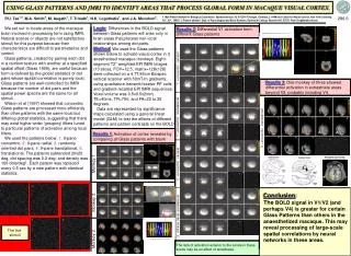
Monkey 2 Monkey 3 USING GLASS PATTERNS AND fMRI TO IDENTIFY AREAS THAT PROCESS GLOBAL FORM IN MACAQUE VISUAL CORTEX. 1. Max-Planck-Institute for Biological Cybernetics, Spemannstrasse 38, D-72076 Tübingen, Germany; 2. HHMI and Center for Neural Science, New York University, N.Y., N.Y. 10003. 3. Present address: Dept. of Psychological and Brain Sciences, Dartmouth College, Hanover N.H. 03755; Peter.Tse@dartmouth.edu P.U. Tse1,3, M.A. Smith2, M. Augath1, T. Trinath1, N.K. Logothetis1, and J.A. Movshon2. 286.6 We set outto locate areas of the macaque brain involved in processing form using fMRI. Natural scenes or objects are not satisfactory stimuli for this purpose because their characteristics are difficult to parameterize and control. Glass patterns, created by pairing each dot in a random texture with another at a specified spatial offset (Glass 1969), are useful because form is defined by the global statistics of dot pairs whose spatial correlation is purely local. Glass patterns are well-controlled for fMRI because the number of dot pairs and the spatial power spectra are the same for all stimuli. Wilson et al (1997) showed that concentric Glass patterns are processed more efficiently than other patterns with the same local but differing global statistics, suggesting that there may exist higher-order 'grouping' filters tuned to particular patterns of activation among local filters. We used the patterns below: 1. 9-pane concentric, 2. 9-pane radial, 3. randomly oriented dot pairs, 4. 9-pane translational, 5. translational. The patterns subtended 20x20 deg, dot spacing was 0.2 deg, and density was 100 dots/deg2. Each pattern was replaced every 0.5 sec by a new pattern with identical statistics. Logic: Differences in the BOLD signal between Glass patterns will arise only in brain areas that process non-local relationships among dot pairs. Method: We used the Glass patterns shown below to activate visual cortex in 3 anesthetized macaque monkeys. Eight-segment T2* weighted EPI fMRI images (13 slices, FOV=12.5, matrix=128x128) were collected on a 4.7T/40cm Biospec vertical scanner with 50mT/m gradients, using quadrature transmit/receive RF coils and gradient-recalled EPI fMRI sequences. Voxel volume was 0.5x0.5x2mm, TE=40ms, TR=750, and FA=20 to 25 degrees. Data are represented by significance maps calculated using a general linear model (GLM) to test the effects of different patterns and pattern contrasts on the BOLD activation. Results 2: Differential V1 activation from different Glass patterns translational x 9 translational x 9 random random concentric x 9 concentric x 9 translational translational radial x 9 radial x 9 # of brain volumes (16 minutes) 1: +concentric -random + - Results 3: One monkey of three showed differential activation in extrastriate areas beyond V2, probably including V4. icon1 icon2 1: +concentric -translational 3: +concentric -translational + - Results 1: Activation of cortex revealed by comparing all Glass patterns with blank + - 2: +concentric -random Monkey 1 Approximate area locations in sample horizontal section + - 1. 2. 3. Conclusion: The BOLD signal in V1/V2 (and perhaps V4) is greater for certain Glass Patterns than others in the anaesthetized macaque. This may reveal processing of large-scale spatial correlations by neural networks in these areas. 2: +9frame transl.-translational 4. 5. + - The five stimuli translational x 9 translational x 9 translational concentric x 9 concentric x 9 translational radial x 9 radial x 9 random random # of brain volumes (16 minutes) The lack of activation anterior to the lunate in these scans may be an effect of anesthesia.