Bacterial Stains
Bacterial Stains. Overview. In our laboratory, bacterial morphology (form and structure) may be examined in two ways: by observing living unstained organisms (wet mount). by observing killed stained organisms.

Bacterial Stains
E N D
Presentation Transcript
Overview • In our laboratory, bacterial morphology (form and structure) may be examined in two ways: • by observing living unstained organisms (wet mount). • by observing killed stained organisms. • Besides being very small, bacteria are also almost completely transparent, colorless and featureless in their natural states. • Microscopy solve the size issue. • Staining can make the structures of bacteria more pronounced. • Since bacteria are almost colorless and therefore show little contrast with the broth in which they are suspended, they are difficult to observe when unstained. Staining microorganisms enables one to: 1: see greater contrast between the organism and the background, 2: differentiate various morphological types (by shape, arrangement, gram reaction, etc.), 3: observe certain structures (flagella, capsules, endospores, etc.).
What is a stain? • A stain (or dye) usually consists of a chromogen and an auxochrome. Reaction of a benzene derivative with a coloring agent (or chromophore) forms a chromogen. The auxochrome imparts a positive or negative charge to the chromogen, thus ionizing it. The ionized stain is capable of binding to cell structures with opposite charges. Example of a dye: Methylene Blue Chloride MBCl MB+ + Cl-
Basic stains (positive stain) are cationic; when ionized, the chromogen exhibits a positive charge. Basic stains bind to negatively charged cell structures like nucleic acids. Methylene blue, crystal violet and carbolfuchsin are common basic stains. • Acidic stains (negative stain) are anionic; when ionized, the chromogen exhibits a negative charge. Acidic stains bind to positively charged cell structures like proteins. Picric acid, eosin and nigrosin are common acidic stains. • Positive stains: Dye binds to the specimen • Negative stains: Dye does not bind to the specimen, but rather around the specimen.
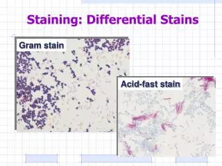
Type of staining in Micro lab 1. Simple stain 2. Differential Stain • Gram stain • Acid fast Stain 3. Special stain • Capsular stain • Endospore stain • Flagellar stain
Part One Simple stain
Simple stain • In this exercise, we will use simple stains to show the general structures of some bacteria. Usually, a single basic stain is used in the procedure. Simple stains do not usually provide any data for identification of the bacterium; they simply make the bacterium easier to see. • To observe basic external structures of cell with bright field scope (cellular morphology)
Method 1. Obtain broth cultures of the bacteria. 2. Using an inoculating loop, remove a loopful of suspension from one of the tubes. Remember to use sterile technique. 3. Smear the bacteria across the center of the slide with the loop. If the bacterial suspension is very thick, add a drop of water and mix the bacteria and the water on the slide.
Method 4. Allow the smear to completely air dry. • Air dry first to prevent lysis (boiling) 5. Heat-fix the smear by quickly passing the slide through a Bunsen burner flame three times. This causes partial melting of the cell walls and membranes of the bacteria, and makes them stick to the slide. Do not overheat the slide as this will destroy the bacteria. Heat Fixing • Kill. • Stops autolysis. • Adherence to slide. • Dye taking 6. Cover the smear with a few drops of one of the stains. Allow the stain to remain for the following periods of time: Carbolfuchsin- 15-30 seconds. Methylene blue- 1-2 minutes. Nigrosin- 20-60 seconds.
7. Gently rinse the slide by holding its surface parallel to a gently flowing stream of water. 8. Gently blot the excess water from the slide with bibulous paper. Do not wipe the slide. Allow the slide to air dry. Observe the slide under the microscope with air and oil lenses. A coverslip is not required. Repeat this process with the other bacteria and stains. Note the differences between the various types of stains and their appearances
What to do??? • DRAW what you see under the microscope • Take not of • Shape • Color • Arrangement • Size of single cells
The simple staining makes it possible to see bacteria clearly, but it does not distinguish between organisms of similar morphology.
Part two Differential stain • gram stain • Acid fast stain
Gram stain • Differential stain (Hans Christian Gram, a Danish doctor ). He developed a new method to stain bacteria so they can be visible in specimen samples. • The most important stain • Differentiate bacteria into two large groups (the Gram Positive and the Gram negative) • Almost all bacteria are described by their Gram stain characteristics. • Based on differences of Cell wall structures
Importance of Gram Stain • This staining method is still valuable today. • It is used in bacterial identification. • It is of great importance in diagnosis of infectious diseases in culture and directly from clinical samples. • For instance, the majority of Gram-positive organisms are susceptible to penicillin, while gram-negative bacteria are resistant to this antibiotics. • It is also valuable to microbiologists, who can plan their culture procedures based on their knowledge of the bacterial forms they have seen in the specimen. • The numerous modifications of Gram’s original method are based on the concentration of the dyes, length of staining time for each dye, and composition of the Decolorizer.
Reagents for Gram Stain • Crystal Violet (purple). • Primary stain; positive stain • Stains cell wall purple • Iodine • Mordant • Combines with primary stain to form an insoluble complex that gets trapped in thicker peptidoglycan layers • Ethanol • Decolorizer • CV-I complex washed out of Gram negative organisms because it cannot be trapped by peptidoglycan layer; flows right through outer membrane • Safranin (pink) • Counterstain • Simple positive stain that provides contrasting dye for decolorized cells (Gram negative) • Stains all cells, but only the negative ones actually appear pink.
When slides are dry, label them as shown: • Examine all slides under oil with the oil-immersion objective.
Artifacts Crystal violet precipitate on epithelial cell: May be confused with Gram positive cocci Crystal violet precipitate crystal on gram stain
Errors during staining • Never ever used old culture. • Time of Decolorizer: • Over: G + see as G -. • Low: G- see as G +. • Time of fixation: • Over: G + see as G -. • Low: no sample on slide.
The acid-fast stain (modified Ziel-Neelsen method). • The acid-fast stain is another differential staining method. • In this case, the target cells are usually members of the genus Mycobacterium. • The cell walls of these bacteria contain an unusually high concentration of waxy lipids, thus making conventional simple stains and Gram stains useless. • The genus Mycobacterium contains two important human pathogens, M. tuberculosis and M. leprae, which cause tuberculosis and leprosy, respectively.
Acid Fast Reagents • Carbolfuchsin (red), a phenolic stain: is the primary stain in the acid-fast test. It is soluble in the lipids of the mycobacterial cell wall. • Heating the specimen, or adding a wetting agent such as Tergitol, increases the penetration of the carbolfuchsin. • Following application of the carbolfuchsin, the specimen is cooled and decolorized with a solution of 3% hydrochloric acid and 95% ethanol (acid-alcohol). • Since carbolfuchsin is more soluble in waxy cell lipids than in acid-alcohol, the acid-alcohol removes the carbolfuchsin from non-acid-fast organisms, but not from acid-fast organisms. Following decolorization, the sample is counterstained with methylene blue which Cannot penetrate mycolic acid; provides contrast to non acid fast cells.
Procedures • Prepare a smear organism and a on glass slides. • Allow the slides to air dry, and then heat fix the organisms. • Apply enough of carbolfuchsin with Tergitol to cover the bacteria. Allow it to set for five minutes. (Kinyoun stain)
Procedures • (Alternate) zielh nelson If Tergitol is not available, apply enough carbolfuchsin to cover the bacteria. Place the slide on a pre-warmed hot plate set on low for 8 minutes. Do not allow the stain to evaporate or Boil. Add additional stain, if necessary. Remove the slide and allow it to cool. • Rinse the slide with acid-alcohol (15-20 sec), drop by drop, just until the alcohol runs clear. • Gently rinse the slide with water.
Apply enough methylene blue to cover the bacteria. Allow it to set for 30 sec. • Gently rinse the slide with water. • Blot (don't wipe) the slide dry with bibulous paper. Allow the slide to air dry. • Examine the slide under oil immersion. Positive organisms will appear pink or red; negative organisms will appear blue.
Under the microscope Acid Fast bacilli (red) Non Acid Fast bacilli (blue) Acid Fast bacilli (red) mixed with non acid fast (blue cocci
Part 3 • Special stains • Capsular stain • Flagellar stain • Endospore stain
Special stain • Emphasize certain cell parts • Some bacteria have characteristic surface structures (such as capsules or flagella) and internal components (e.g., endospores) that may have taxonomic value for their identification. When it is necessary to demonstrate whether or not a particular organism possesses a capsule, is flagellated, or forms endospores, special staining techniques must be used.
Bacterial endospores • Resting structures formed by some bacteria for survival during adverse environmental conditions (nutrient limitation or extreme environments) • The endospore is a highly resistant differentiated bacterial cell that are highly resistant to heat, boiling and drying out and are difficult to destroy • Endospores can remain dormant indefinitely ((not reproductive), but germinate quickly when the appropriate trigger is applied • Metabolically inactive • Stable for years • Endospores differ significantly from the vegetative , or normally functioning, cells • Formed by Gram-positive bacteria • (e.g. Bacillus, Clostridium)
Staining procedures • Malachite green is the primary stain .which is placed on blotting paper over the smear gently heating over a warm water bath to penetrate the spore coat. • The bacteria aredecolorized with water.leaves theendospores green as the stain is driven into the endospore . The malachite green is washed out of the vegetative cells with the water. • It is then counterstainedwith safranin. • Do not allow the stain evaporate. to prevent formation of metallic sheet
Problems Interpreting Endospore Stain • It should be noted that any debris on the slide can also take up and hold the green stain. Everything that ends up green on the slide is not necessarily an endospore. Endospores are small and typically oval. Large or irregular globs of green on the slide may be artifacts. • Acid-fast cells, such as members of MycobacteriumandNocardia have waxy molecules in their cell wall that will take up and retain the malachite green stain when subjected to the endospore staining process. • Endospores killed when dry heat is applied at high temperatures or for long periods, by steam heat under pressure (in the autoclave), or by special sporicidal (endospore-killing) disinfectants.
What is Capsule? • Capsules are structures composed of carbohydrate or glycoprotein that lay outside of an organism's cell wall and thus are in direct contact with the environment. Many bacteria produce capsules under the right conditions.
Functions of a capsule • Protect the cell from desiccation (drying) • Protect the cell from phagocytes (being engulfed by white blood cells) • Provide a food reserve when certain organic compounds are in excess. • A virulence determinant of pathogenic microbes • They serve as binding or adhesion agents for sticking cells together and/or to a surface such as a rock in flowing stream or a tooth