Download

1 / 17
200 likes | 513 Vues
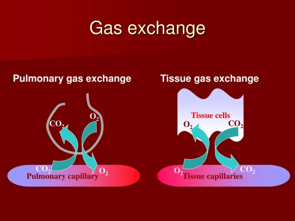
Gas Exchange and Transport. Gas Exchange and Transport. The driving force for pulmonary blood and alveolar gas exchange is the Pressure Differential – The difference between the partial pressure of a gas (O 2 or CO 2 ) above a fluid and dissolved in fluid (alveoli or blood).

E N D
Gas Exchange and Transport The driving force for pulmonary blood and alveolar gas exchange is the Pressure Differential – The difference between the partial pressure of a gas (O2 or CO2) above a fluid and dissolved in fluid (alveoli or blood)
Gas Exchange and Transport Pressure Differential Fig 13.1
Gas Exchange and Transport • Henry’s Law: • The rate of gas diffusion into a liquid depends on: • Pressure differential between the gas above the fluid and gas dissolved in fluid • Solubility (dissolving power) of the gas in the fluid CO2 highly soluble
Gas Exchange and Transport Saturation with water vapor - lower PO2 Constant loading and unloading of CO2 and O2 FRC necessary to prevent swings in CO2 and O2 concentration in alveoli PO2 – 100 mm Hg:regulates breathing and 02 loading of Hb PCO2 – 40 mm Hg:chemical basis for ventilatory control via respiratory center Fig 13.2
Gas Exchange and Transport Time Required for Gas Exchange Fig 13.2 Capillary transit time is ~0.75 s During maximal exercise, capillary transit time is ~0.4 s Gas exchange during maximal exercise not a limiting factor
Gas Exchange and Transport Time Required for Gas Exchange Fig 13.2 Pulmonary disease impacts this process: 1. Thicker alveolar membrane 2. Reduced surface area Fick's Law-Gas diffuses at rate proportional to: Tissue thickness (inversely) Tissue area (directly)
Gas Exchange and Transport • O2 Transport: • Dissolved oxygen in blood only sustains life for about 4 seconds (0.3 mL O2 / dL) • Small amount establishes PO2 which regulates breathing and oxygen loading of hemoglobin
Gas Exchange and Transport • O2 Transport: • Hemoglobin (Hb) – Protein in red blood cells that transports 02 bound to iron • Each Hb has 4 iron atoms (can bind 4 O2) • Hb transports 19.7 ml/dL (vs 0.3 ml/dL - plasma) • (65 x that in plasma) Fig 13.3 Anemia: Low iron in red blood cells results in low oxygen carrying capacity
Gas Exchange and Transport Oxyhemoglobin dissociation curve: Fig 13.4 Describes Hb saturation with O2 at various PO2 levels 100 mm Hg: 98% saturation 60 mm HG: decline in % saturation 40 mm HG: 75% of O2 remains with Hb - 5 ml delivered to tissues Athletes?
Gas Exchange and Transport Fig 13.4 • Bohr effect – • Increased blood acidity (lactic acid), temperature, CO2 causes downward shift to the right • Facilitates dissociation of O2 from Hb • No effect on capillary blood Hb-O2 binding
Gas Exchange and Transport Oxyhemoglobin dissociation curve: Fig 13.4 • Myoglobin: • Intramuscular O2 storage protein • Transfers O2 to mitochondria when PO2 falls • At 40 mm Hg, Mb 95% saturated with O2 • No Bohr effect occurs with myoglobin
Pulmonary Ventilation Ventilatory Control – How does our body control rate and depth of breathing in response to metabolic need Medulla – Inspiratory neurons activate diaphragm and intercostals Expiratory neurons activated by passive recoil of lungs *Mechanisms maintain constant alveolar and arterial gas pressures Fig 14.1
Pulmonary Ventilation 1. At rest, chemical state of the blood controls ventilation PO2, PCO2, acidity (lactate), temperature PO2 – no effect on medulla (peripheral chemoreceptors detect arterial hypoxia, altitude) PCO2 – most important respiratory stimulus to medulla at rest Fig 14.2
Pulmonary Ventilation 2. During exercise, no single mechanism explains increase in ventilation (hyperpnea) • Neurogenic Factors: • Cortical: Motor cortex stimulates respiratory neurons to increase ventilation • Peripheral: Mechanoreceptors in muscles, joints, tendons influence ventilatory response • Peripheral chemoreceptors become sensitive to CO2, H+, K+, and temperature during strenuous exercise
Pulmonary Ventilation Phases of Ventilatory Response During Exercise: I. Neurogenic – central command, peripheral input stimulates medulla Fig 14.4 II. Neurogenic – continued central command, peripheral chemoreceptors (carotid) III. Peripheral - CO2, H+, lactate (medulla), peripheral chemoreceptors Rapid rise Slower exponential rise Steady state ventilation Abrupt decline Recovery – removal of central, peripheral, chemical input