Download
1 / 105
1.09k likes | 1.6k Vues
Semiolog y of immune system. Department of pediatrics. No t i o n. Immune system reunites the organ s , tissues and cells , which ensure the defense of h uman organism against the genetically strange substances (antigen s ) by exogenous and endogenous origin . Immune system.

E N D
Semiologyof immune system Department of pediatrics
Notion Immune system reunites the organs, tissues andcells, which ensure the defense ofhuman organism against the geneticallystrange substances (antigens) by exogenous and endogenous origin.
Immune system Two possibilities of IS response manifesting were distinguished for to understand how the organism succeedsto put up resistance to the external medium factors (infectious agents) : elimination ofpathogenic agents with the nonspecific means of IS response or producing of some cellular and molecular componentsfor adopting the pathogenic agent (specific means).
Physiologic importance The function of IS consists in the Ag identifying and generation of specific response – synthesisofantibodies, accumulation of sensibilized lymphocytes, which will neutralized, destroyedand eliminated the Ag from organism.
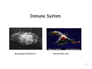
Components of immune system 1. Lymphoid organs: a) Central b) Peripheral 2. Humoral components a) Nonspecific b) Specific 3. Cellular components a) Nonspecific b) Specific
Central lymphoid organs The central lymphoid organs (primary) are represented by: hematogene marrow thymus
Central lymphoid organs The central lymphoid organs have a major significance inimmunitybecause they represent theplace oflymphopoiesis. At their level, the cellular components of IS: B lymphocytes and T lymphocytes, aredifferentiatingfrom derived precursors originated from stem cell, proliferatingand se maturating in functional cells.
Hematogene marrow Hematogene marrow is localizing in the trabeculae of osseos spongious tissue fromlong bones epiphyses, from flat bones and from short bones. From the histologic point of view, it is formed from stroma, blood, lymphatic vesselsand nerves. The stroma is formed from conjunctive reticular cells, forminga network where are sitting the stem cells. Here the B lymphocytes are differentiating.
Thymus The thymus is sitting retrosternally. From the anatomic point of view, the thymus is formed from two lobes connected by isthmus. Each lobe is formed from lobules delimitedby conjunctive septa whichproceed fromthe capsula covering the entire organ. Two zones are described in the thymic lobule: corticaland medullar. The thymic cells are lymphocytes, epithelial and non-epithelial cells. The process of differentiation is developing under the influence of local hormones (thymosine, thymopoietine, thymuline) secretedespecially by subcapsular and subtrabecular epithelium.
Thymus The thymus is forming at the end of first month of intrauterine development. It is positioned retrosternally. It is covered by conjunctive tissue capsulawhichseparates the organ in lobules. Each lobulehas cortical and medullar layer. The cortical layer contains T-lymphocytes, and epithelial cells of medullar layer form the Gassale corpuscles.
Thymus The thymus releases in the systemic circulation T-lymphocytes, hormones (thymosine, thymopoietine, thymic factor etc.) whichregulates the proliferationand differentiation of lymphocytes. The thymus achieves the maximal degree of development in early childhood. In the period of 3 – 13-15 yrsthe stabilizationof gland mass has place, and ulterior it involutes. The cortical layer becomesmore poor in T-lymphocytes, the Gassale corpuscles from medullar layer disappear, thesestructures beingreplacedwith conjunctiveand adipose tissue.
Thymus In rare cases the physiologic involutionis not producing. These clinical situationsusuallyare associatedwithdiminishing of GCS secretionby cortical layer of suprarenal glands. Such patientsare more receptiveto intercurrent infections, have increased risc forneoplastic processes appearance.
Peripheral lymphoid organs Peripheral (secondary) lymphoid organs are represented by: Spleen Lymph nodes Lymphoid tissue from mucosae
Spleen The spleen is situatedin the left superior part of abdomen. It has exterior capsulafrom conjunctive tissuewhichsendsin parenchymaprolongationsforming trabeculae. Thesetogetherwith the reticular cells network form a support for agreat lot of cells. Two types of tissue enterin the spleen structure : the tissue responsibleforgrown old blood cells destruction and urgent generation of new erythrocytes, plateletsand granulocytes (red pulp) and tissue populatedbyimmune competent cells (white pulp).
Spleen Spleen white pulp is a lymphoid tissue situatedin two zones: T-dependent zone, situatedaround central arteriolaand B-dependent zone, whichsurroundsthe T-zone, as a muff. In B-zone thecells are organizedinprimary follicles (non stimulated) and in secondary follicles (stimulated). At the periphery of B-zone, to the exterior,the splenic macrophages are sitting.
Lymph nodes The lymph nodes are oval formations by different dimensions, situated in theconfluence placeofgreat lymph vessels.
Lymph nodes The forming of LN begins in the 2-ndmonth of intrauterine developmentand is finishing in thepostnatal period. In new-born the LN capsula is very thin and fine, the trabeculae are weakly differentiated, and their palpationis difficult. LN are soft and they are includedin good developed adipose tissue at this age. At 1 year age theLNcan be palpated at the majority ofchildren. Parallel with their growing their differentiation has place.
Lymph nodes In the 3 yrs age the capsula is good formed. At 7-8 yrstheforming of trabeculae in the interior ofLN begins. At 12-13 yrs the structure of LNisdefinitized, being good differentiated all theirstructures: capsula, trabeculae, follicles, sinuses.
Lymph nodes In the puberty period the LN growing is finishing, and sometime evenpartially regresses. The maximal number of LN is achieved around10 years age. The mature has approximatively 460 LN, having total mass by1% from corporal mass (500 – 1000g).
Lymph nodes Each lymph node (LN) is covered at surface with conjunctive tissue capsule, and in interior contains lymphoid tissue. The lymphoid tissue of LN isseparated in two layers: corticalandmedullar. Cortical layer isformedfromfollicles– conglomeratesof B lymphocytes. Inparacorticallayer the T-lymphocytes are placed Inmedullarlayer the plasmocytes, secreting immunoglobulins are placed.
Lymph nodes LNareplaced in groups, throughthem the lymph drainage from distinct anatomic zones has place. Due to theirstructure and localization theLN have a roleofbarrierin the pathway of infection spreading, preventing its generalization. LNfilter the particleswith antigenic properties, and the lymphocytesand plasmocytesfrom LN ensure the synthesis of antibodies. The lymphoid system of respiratoryand digestive tract ensures the good functioning of local immunity at the level of their mucosa.
Lymph nodes The reaction of LN at different stimuli, especially infectiouscan be observed from 3 months age. In 1-2 yrs age children thebarrier function of LN is not good expressed, therefore in this age the infections easily are generalized (septicemia, meningitis, generalized forms of TBC). Immaturity of lymphoid system at the level of digestive tubepredisposes thesuckling babiesat intestinal infectionsand allergization of organism on enteral pathway.
Lymph nodes In the antepreschool age theLNare still good structuredand can serveas mechanicalbarrier against the infection spreading. In this age the lymphadenites, including these purulent and caseous (TBC) are frequent. In the age of 7-8 years the LNbecome functional and can suppress the infection through immunologic mechanisms.
Lymph nodes Occipital– placedon occipital tuberozities – they collect the lymphfrom the scalp skinand cervical posterior region. Mastoidian– placedin region of mastoid apophises, retroauricular – placed postrior fromears pavilion – both groups collect the lymphfrom medium ear, external auditive conduct, ears pavilion, paraauricular teguments. Submandibular – placedunder inferior bundlesof mandiba – collect the lymphfrom face skin andgums mucosa. Mentoniar– placedone self bilaterallyin the region ofchin – collect the lymphfrom the inferior lip skin, mucosa of gumsand from inferior incissives.
Lymph nodes Anterior cervical and tonsilar – placed anterior from the sternocleidomastoidian muscle, in superior cervical triangle– collect the lymphfrom face teguments, from parathyroid gland, nasal, faringealand buccalmucosa. Posterior cervical – placed posterior fromthe sternocleidomastoidian muscle, anterior fromtrapezium muscle, in cervical superior triangle – collect the lymphfrom cervical region and partiallyfrom larynx. The LNfrom above mentioned groups are frequently cataloguedas a unique group – cervical LN.
Lymph nodes Supraclavicular– placedin supraclavicular fossa – collect the lymphfrom superior partofthorax, pleural domes and pulmonary apexes. Subclavicular– placedin infraclavicular fossa – collect the lymphfrom the thoracic walland pleura. Axillary – placed în axillary fossa – collect the lymphfrom superior members (exceptionfingers V, IV, III and palm). Thoracic– placedon the inferior margin ofbig pectoral muscle – collect the lymphfrom thoracic wall teguments, parietalpleura, partiallyfrom lungs and mammal glands.
Lymph nodes Cubital – placed in the cubital regionat the level of bicepstendon – collect the lymphfrom V, IV, III fingers and palm. Inguinal – placedonline of inguinal ligament– collect the lymphfrom the teguments of inferior member, hypogastrium, buttocks, anal region, perineum, genital organs. Popliteal– placedin the popliteal fossa – collect the lymphfrom the teguments ofleg.
Lymph nodes The knowledge of LN localizationand of their draining zoneshas a major role in theidentifying ofinfectiongateway, especially in the case when the modificationsof infection gateway are minimal oreven absent, but regional LNwill reactin the all cases.
The semeiology of LN affection Complaints. In the case of LN marked enlargement, the child or the parents can remark this modificationand can present the respective complaints. In the case of lymphadenitis thechild canaccusepain, tumefactionand hyperemiaat the level of affected LN.
Semeiology of LN affection Inspection. Only LN which are placed superficially and are very enlarged in volume (lymphogranulomatosis, infectious mononucleosis) can be observed. In lymphadenitisthe hyperemia of tegumentscovering theinvolved LN, which usually is tumefied and painful at palpation can be observed.
Semeiology of LN affection Palpation. At palpation the LNthe following Characteristics are appreciated: Dimension – usually theLNhave a diameter around 0,3-0,5 cm (pea seed). The dimension of LNisgradated as the following: I degree – dimension of millet seed II grad – dimension oflentil seed III grad – dimension of pea seed IV grad – dimension ofhorse bean V grad – dimension of peanut VI grad – dimension of pigeon egg. Increasing of LN dimension poate can be isolated or in group, symmetrical or unilateral.
Semeiology of LN affection Number If in each group 3 or less LN are palpable – they are considered solitary, If in some group more than 3 LNare palpated – they are considered multiple.
Semeiology of LN affection Consistence Can be soft, elastic, hard. It depends from the oldness and nature of process: in chronic pathology the LN are hard, in the case of recent affectionthey have soft consistence. Physiologically the consistence of LN is elastic.
Semeiology of LN affection Mobility – usually the LN are mobile. Reportwith teguments, adipose subcutaneous tissue, other LN. Physiologically the LN don’tconnectwith adiacent tissuesand not between them. Sensibility. LN are painless at palpation. The presence of pain at palpation indicates an acute inflammatory process at the level of LN.
Semeiology of LN affection The palpation of symmetrical LN isperforming with both hands concomitantly. (exception – cubital LN). In healthy children until 3 groups of LN are palpable. In physiological conditions the The chin, supra- and subclavicular, thoracal, cubital and poplitealLN are not palpating.
Semeiology of LN affection The LN can be considered normal, if: their dimension does notexceed the dimension of pea seed, are solitary, by elastic consistence, mobile, don’t adherebetween them and adjacent tissues, are not painful.
Semeiology of LN affection For the performing of certain diagnosis, the clinical examination ofLN is supplementedat necessitywith special paraclinicalexaminations: puncture of LN, biopsy of LN, lymphography.
Semeiology of LN affection In children the modifications of LN both local andgeneralized are frequently observed. Local/regional enlargementof LN accompanies the suppurative processesat the level of teguments: folliculites, pioderma, furunculosis, miliary multipleabscesses , infected sores, hydroadenitis etc.
Semeiology of LN affection Lymphadenopathy – enlargement of LN in dimensions, sometimes with modification of their consistence. Polyadenia – increasing of number of palpable LN.
Semeiology of LN affection In diphtheria, scarlet fever, tonsillitis the cervical LN react. În felinosisthe cubital and/or axillary LN are modified.
Semeiology of LN affection TBC of peripheral LN usuallyislimiting at the affection of one group ofLN, most often cervical. The LN represent a voluminous, hard, painless conglomerate (packet). They are adherent between them and adjacent tissues. They have tendency of evolution to the caseous necrosis, with forming of local fistula, after that the „ stellated scars” remain.
Semeiology of LN affection In theinfectious mononucleosis all groupsof peripheral LN are affected, but the affection of cervical posterior LN predominates. In rujeolathe occipital LN are affected. For rubeola the diffuse LN affection, more pronounced in cervical, occipital and axillar LN groups is characteristic.
Semeiology of LN affection In adenoviral and paragrippal infection the anterior and posterior cervical and occipital LN are predominantly involved. In mumps the auricular LNare palpating as firm formations which are placed superiorly to parotidian tumefiated glands. In toxoplasmosismost frequently thegroups of cervical, axillary, inguinal LN are affected. The LN have the dimensionsof a nut, sometimes can have a form of pockets, but each node can be palpated separately.
Semeiology of LN affection In HIV/AIDS thegeneralized lymphadenopathyis a precociousand constant symptom. The diameter of LN is 2-3 cm, the contur net delimitated, hard atpalpation, not adhere between them and not to adjacent tissues, usuallyare sensibleat palpation, sometimes painful.
Semeiology of LN affection Lymphosarcoma ismanifesting by affectionof isolated LN group, usually cervical or supraclavicular. LNare very hard, painlessatpalpation, the local signs of inflammation are absent. The generalized lymphadenopathy can be present in a lot of acute or chronic infectious pathologies and in noninfectious pathologies.
Semeiology of LN affection The diffuse diseases of conjunctive tissue can be around the noninfectious causes of generalized lymphadenopathy. Lymphogranulomatosisas a rule beginswith peripheral LN affection, more frequently cervical or/and mandibular. In the same timewithdisease evolution the LN grow in conglomerates. LN are painless, atpalpationcreates a sensation of „sackofpotatoes”. The diagnosis is confirmed by the hystologic examination of LN (Berezovski-Sternberg cells). In ALL (acute lymphoblastic leukaemia) all groups of LN are rapidly growing in dimensions. They are soft and painless atpalpation.
Lymphoid tissuesassociated to mucosae The lymphoid tissues associated to mucosae also enter in the category of secondary lymphoid organs. These can be: good individualized anatomically structures or lympho-epithelial diffuse structures.