Download
1 / 10
100 likes | 479 Vues
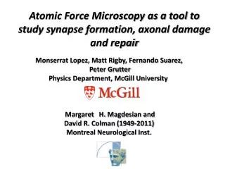
Atomic Force Microscopy as a tool to study synapse formation, axonal damage and repair. Monserrat Lopez, Matt Rigby, Fernando Suarez, Peter Grutter Physics Department, McGill University Margaret H. Magdesian and David R. Colman (1949-2011) Montreal Neurological Inst. .

E N D
Atomic Force Microscopy as a tool to study synapse formation, axonal damage and repair Monserrat Lopez, Matt Rigby, Fernando Suarez, Peter Grutter Physics Department, McGill University Margaret H. Magdesian and David R. Colman (1949-2011) Montreal Neurological Inst.
Magnetic reversal Molecular electronics Quantum dots Interfacing to living neurons Biochemical sensors MFM with in-situ field UHV AFM/STM/FIM, AFM/STM/SEM 4K, 8T and 50mK, 16T AFM AFM + SNOM + patch clamp + single photon fluorescence + TIRFM Cantilevers and electrochemical AFM SPM applied to nanoelectronics: the Grutter Research Group www.physics.mcgill.ca/~peter
axon spine Synapse formation is accompanied by change in mechanical properties Left: Hippocampal neuron imaged with an AFM probe. Right: Corresponding stiffness maps (bright is soft, dark is stiff). Spines appeared soft relative to the dendrite shafts, where stiff patches or fibers were identified (small arrows). Spine shapes were irregular, often exhibiting small surface protrusions (arrowheads). Axons were not observed in close proximity to the soft spines. Ben Smith et al., Biophys. J. 92, 1419 (2007)
Studying Axonal Degeneration by AFM • Advantages • live imaging during gradual injury • precise control of injury parameters (positioning and force determinations in the sub-nanoscale) Before Compression Recovery Deformation Increased deformation Degeneration Hippocampal DRG
Results Using the AFM as an imaging tool we can follow the morphological response of axons to injury. Using the AFM in force spectroscopy mode to cause gradual damage to axons we can follow the response of different axonal components to injury. Hippocampal axons can support 100 ± 50 Pa for 10 minutes while DRG axons resist up to 500 ± 200 Pa for 30 minutes. Axonal rupture - optical labeling of components allow determination of failure mechanism: axonal stiffness decrease due to disrupting microtubules and degeneration due to disruption of mitochondrial transport. mitochondria tip Axon Magdesian et al., 2012
Forming a synapse with a functionalized bead attached to an AFM tip: controlling synapse location and time! TEM and SEM indicate that structure of bead-induced synapse is identical to a natural synapse. Attach bead to AFM tip! Allows recruitment time of various (labelled) proteins to be quantified. Allows extraction of functioning neuronal filaments! Observation: Rapid assembly of functional presynapticboutons triggered by adhesive contacts with Poly-D-Lysin coated beads attached to AFM tip (circle above). Control: uncoated beads – no recruitment! Stable synapes formed Recruited!
“Neurite” (S) formation observed upon pulling the PDL coated bead away from an axon. Neurons labeled with synaptophysin-GFP. We have observed “neurites” as long as 50um. These neurites contain tubulin,actin, bassoon and synaptophysin. Using the AFM to repair axons Images from Fernando S. Sanchez unpublished
Using the AFM to repair axons Detector Model Laser Cantilever PDL-Bead 1 2 3 20 min. contact Lift cantilever 20 min. contact Lift cantilever
Using the AFM to repair axons 2 3 2 1 2 1 20 min contact lift cantilever 20 min. contact lift cantilever with axon 1 pulling “neurite” with axon 2 Problem: the neurite is not dettaching from the PDL-coated bead