Download
1 / 2
20 likes | 113 Vues
Investigating the interaction of TNF-α with HMGB-1 gene in influencing immune response through molecular methods like ligation, transformation, and Western blotting. Analysis of cytokine-induced inflammation and its regulatory potential for immune system functions.

E N D
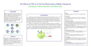
The Effects of TNF-α on the Proinflammatory HMGB-1 Response Zoë Kabelac, Theresa Alexander and Andrea Fisher Methods Ligation HMBG-1 was added into an E.coli plasmid, using HindIII and XbaI restriction enzymes Transformation The amount of recombinant DNA in the plasmid was amplified by inserting the DNA into an E.coli bacterial cell through a calcium chloride treatment and a heat shock. DNA Preps (Mini & Midi) Mini: In an overnight culture, bacteria was grown on a small scale. The bacteria was then purified and the recombinant plasmid was taken out of the cells by washing it through a column with various buffers. Then it was run through a gel to verify that the cells were transformed with the recombinant plasmid containing the HMGB-1 gene. Midi: Bacteria was grown in an overnight glycerol stock on a large scale. The bacteria was then purified and the recombinant plasmid was taken out of the cells and run through a gel to once again verify the HMGB-1 gene was present. Enzyme Digestion and Analysis The DNA sample was digested with XbaI and HindIII enzymes and run through a gel to verify that the cells contained the HMGB-1 gene. Transfection FuGene was added to the recombinant DNA to assist in the smooth transition across the phospholipid bilayer of the eukaryotic cell. Western blotting In preparation for the Western Blotting the cells were lysed in order to obtain the HMGB-1 protein and the results are printed on X-ray film. The Western Blot was used to detect the amount of HMGB-1 protein present in the cells. Immunofluorescence Two different antibodies were used to detect the presence of the HMGB-1 protein. The HMGB-1 protein was tagged with a fluorescent tag. Hypothesis If TNF-α is introduced into a eukaryotic cell containing HMGB-1, then the immune response will be greater than if it were just HMGB-1 alone, because TNF-α is a known mediator of immune response. When HMGB-1 moves out of the cell, it will call the immune cells, such as macrophages to the cell, triggering surrounding cells not just immune cells to mount an immune response. Then the macrophages will release TNF-α in response, therefore increasing the inflammation and creating a synergistic effect between the HMGB-1 and TNF-α. Introduction Inflammation is usually advantageous; it helps the body signal antibodies to fight infections, antigens or foreign bodies. If uncontrolled however, it can damage more tissue than it salvages. This is what occurs in the case of inflammation in the brain, known as encephalitis. If inflammation is unregulated it will damage the surrounding tissue because the inflammation will obstruct the oxygen flow to neighboring tissues. Inflammation is a topic of interest because understanding how and why inflammation occurs is a key component in the regulation of an immune response. It is beneficial to understand the relationship between cytokines and inflammation because in cases like encephalitis, inflammation needs to be suppressed, and in others it needs to be induced. HMGB-1 is an essential protein that assists in the inflammatory response. Usually it acts as a transcription cofactor, but when a cell is under stress or going through necrosis, the HMGB-1 proteins migrate out of the nucleus into the cytoplasm. From the cytoplasm it then moves to the outside of the cell, triggering antibodies from surrounding cells. TNF-α is a cytokine which has a primary role of regulating immune cells. TNF-α is also capable of stimulating apoptotic cells, to induce inflammation, and inhibiting tumorigenesis and viral replication. For TNF-α to be effective, it must be within an optimal range. If there is an overproduction of TNF-α, it can be deadly to neighboring cells; however, if there is too little produced, TNF-α will be ineffective in containing and fighting off infection. Abstract Understanding the relationship between cytokines and inflammation is vital in the comprehension of how and why inflammation occurs and how it can be regulated. Inflammation occurs naturally in reaction to a stimulant that your body perceives as harmful. Researching the effects of cytokines, such as TNF-α, on the HMGB-1 gene and their affects on the immune response will enable the suppression and induction of inflammation.
Conclusion Gel Electrophoresis: In the first gel, a large amount of DNA was apparent near the wells, suggesting the DNA was in large clumps. Since restriction enzymes were added, a faulty result came of this because the HMGB-1 gene was not cut out of the plasmid. In the second attempt, large fragments were seen near the wells in addition to a small fragment near the 650 base pair marker. This shows that the HMGB-1 gene was in the plasmid and was successfully digested out. Immunofluorescence: At the 0 minute mark, the red dye was seen darker in uniform shapes, suggesting the HMGB-1 protein was being expressed in large amounts and was defined in the cytoplasm. There were more punctate spots indicating the HMGB-1 protein was being expressed in the nucleus. At the 40 minute mark, the red dye was faded and not as clearly defined. The staining was more diffuse, suggesting the HMGB-1 protein was dispersing throughout the cell, moving toward the outside. Since the coloring was lighter, it insinuates that the protein had moved out of the cell and into the extracellular fluid, which was washed away. At the 60 minute mark, the red dye was the lightest and most diffuse staining, suggesting the protein continued to move away from the nucleus and toward the outside of the cell. Western blotting: There was too much background, making it difficult to interpret specific proteins. This could have been caused by something precipitating out and binding to the filter. Therefore, the amount of protein being expressed over an extended period of time could not be determined. • Objectives • Analyze the effect TNF has on the position of the HMGB-1 protein • Understand how to regulate immune responses Results Gel Electrophoresis: The gel in figure 1 shows the DNA was purified and present, but it was not digested correctly because the HMGB-1 was not visible by the 650 base pair marker. In the second attempt, indicated in Figure 2, the DNA was apparent on the gel and the HMGB-1 gene was visible by the 650 base pair marker. Immunofluorescence: The HMGB-1, which was dyed red, was evident in condensed, dark clumps within the cells. By the 60 minute time mark, the red stain was lighter and not as condensed. Western Blot: (shown below) The results came back inconclusive because there was too much background on the film, making it unable to determine how much protein was being expressed. Bibliography Davidson College. TNF-alpha. 2 July 2009 <http://www.bio.davidson.edu/COURSES/Immunology/Students/spring2000/wolf/tnfalpha.html>. Harris, Helena Erlandsson, and Angela Raucci. “Alarmin(g) News about Danger: Workshop on Innate Danger Signals and HMGB1.” EMBO Reports: 774-8. 2 July 2009 <http://www.nature.com/embor/journal/v7/n8/full/7400759.html>. Tracey, Kevin J. Rev. of Physiology and Immunology of the Cholinergic Antiinflammatory Pathway. The Journal of Clinical Investigation 117.2: 289-94. Tracey, Kevin J, and Anthony Cerami. “Tumor Necrosis Factor.” Annual Review of Medicine 45 (1994): 491-503.