Download
1 / 1
10 likes | 111 Vues
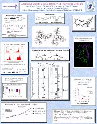
Thioredoxin-targeted Experimental Anti-tumour Quinols Exert Cytotoxicty Following Activation of Apoptosis Signal-regulating Kinase 1. Charles S Matthews, Tracey D Bradshaw, Eng-Hui Chew, Thilo Hagen and Malcolm FG Stevens. Mechanism of Action: Inhibition of Thioredoxin.

E N D
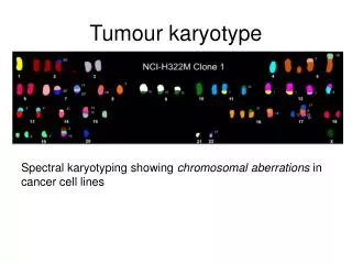
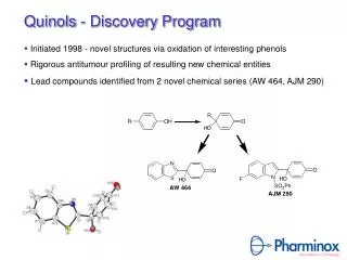
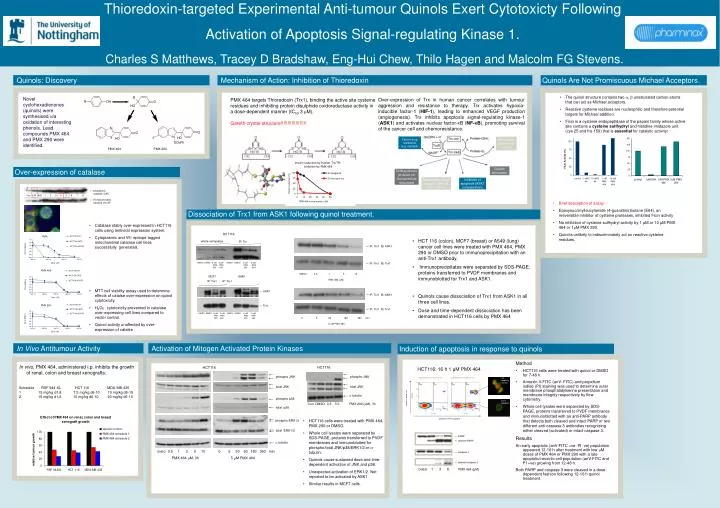
Thioredoxin-targeted Experimental Anti-tumour Quinols Exert Cytotoxicty Following Activation of Apoptosis Signal-regulating Kinase 1. Charles S Matthews, Tracey D Bradshaw, Eng-Hui Chew, Thilo Hagen and Malcolm FG Stevens. Mechanism of Action: Inhibition of Thioredoxin Over-expression of Trx in human cancer correlates with tumour aggression and resistance to therapy. Trx activates hypoxia-inducible factor-1 (HIF-1), leading to enhanced VEGF production (angiogenesis). Trx inhibits apoptosis signal-regulating kinase-1 (ASK1) and activates nuclear factor-KB (NF-KB), promoting survival of the cancer cell and chemoresistance. PMX 464 targets Thioredoxin (Trx1), binding the active site cysteine residues and inhibiting protein disulphide oxidoreductase activity in a dose-dependent manner (IC50 3 μM). Gareth crystal structure!!!!!!!!!!!!!!!!!!! PMX 464 PMX 290 Cancer drug resistance (e.g. cisplatin) Antioxidant (removal of H2O2) Growth stimulation DNA synthesis (H donor for ribonucleotide reductase) Transcription factor regulation (NF-kB, AP-1, Hif1) Inhibition of apoptosis (ASK1 complexation) ← cytoplasmic catalase (CAT) Con EV1 EV2 1 2 3 4 5 6 7 8 ← V5 mitochondrial catalase (mCAT) Identification of Prx-1 as Quinol target ● Quinol, immobilised onto aminoalkyl agarose beads, bound Peroxiredoxin-1 (Prx-1) within HCT 116 cell lysates (A). ● HCT 116 lysates expressing Prx-1-Flag were incubated with beads coupled, or uncoupled to Quinol. Western Blot (anti Flag 1o Ab) detected Prx-1-Flag bound to Quinol coupled beads (B). A. HCT116 whole cell lysates IP: Trx 175 IP: Trx1 IB: ASK1 83 62 IP: Trx1 IB: Trx1 DMSO DMSO 5 M 5 M DMSO DMSO 5 M 5 M PMX PMX PMX PMX 290 464 290 464 47.5 DMSO 0.5 1 5 10 PMX 464 (mM) MCF7 A549 IP: Trx1 IB: ASK1 IP: Trx1 IP: Trx1 32.5 ASK1 IP: Trx1 IB: Trx1 B. Prx-1 25 0 5 60 180 360 min 5 mM PMX 464. Quinol - + Beads + + Prx-1- Flag + + Trx1 16.5 DMSO DMSO 5 M 5 M DMSO DMSO 5 M 5 M PMX PMX PMX PMX 290 464 290 464 Prx-1 Flag Conclusions ● PMX 464 targets the active site cysteine residues of Trx, inhibiting Trx signalling ● PMX 464 fails to inhibit cysteine protease activity ● PMX 464 triggers dissociation of ASK-1 from Trx, initiating JNK and P38 activation ● PMX 464 inhibits HIF signal transduction and NF-kB activation ● Redox protein Prx-1, which possesses a role in H2O2-mediated signalling has been identified as a molecular Quinol target. HCT116 HCT116 Late apoptotic/necrotic total JNK phospho JNK phospho JNK total JNK a tubulin Early apoptotic phospho p38 Propidium Iodide (red) total p38 annexin V-FITC (green) phospho ERK1/2 PARP cleaved PARP total ERK1/2 caspase 3 a tubulin DMSO 0.5 1 3 5 10 0 5 30 60 180 360 min PMX 464 mM, 3h 5 mM PMX 464 cleaved caspase 3 DMSO 1 3 6 PMX 464 (μM) Con DMSO 0.5 5.0 PMX 290 (mM), 3h Quinols: Discovery Quinols Are Not Promiscuous Michael Acceptors. • The quinol structure contains two , unsaturated carbon atoms that can act as Michael acceptors. • Reactive cysteine residues are nucleophilic and therefore potential targets for Michael addition. • Ficin is a cysteine endopeptidase of the papain family whose active site contains a cysteine sulfhydryl and histidine imidazole unit (cys 25 and his 159) that is essential for catalytic activity: Novel cyclohexadienones (quinols) were synthesised via oxidation of interesting phenols. Lead compounds PMX 464 and PMX 290 were identified. Over-expression of catalase • Brief description of assay • Epoxysuccinylleucylamide (4-guanidino)butane (E64), an irreversible inhibitor of cysteine proteases, inhibited Ficin activity • No inhibition of cysteine sulfhydryl activity by 1 μM or 10 μM PMX 464 or 1mM PMX 290. • Quinols unlikely to indiscriminately act on reactive cysteine residues. Dissociation of Trx1 from ASK1 following quinol treatment. • Catalase stably over-expressed in HCT116 cells using lentiviral expression system. • Cytoplasmic and V5- epitope tagged mitochondrial catalase cell lines successfully generated. • MTT cell viability assay used to determine effects of catalse over-expression on quinol cytotoxicity. • H2O2 cytotoxicity prevented in catalase over-expressing cell lines compared to vector control. • Quinol activity unaffected by over-expression of catalse • HCT 116 (colon), MCF7 (breast) or A549 (lung) cancer cell lines were treated with PMX 464, PMX 290 or DMSO prior to immunoprecipitation with an anti-Trx1 antibody. • Immunoprecipitates were separated by SDS-PAGE, proteins transferred to PVDF membranes and immunoblotted for Trx1 and ASK1. • Quinols cause dissociation of Trx1 from ASK1 in all three cell lines. • Dose and time-dependent dissociation has been demonstrated in HCT116 cells by PMX 464 HCT116 ASK-1 Trx1 In Vivo Antitumour Activity Activation of Mitogen Activated Protein Kinases Induction of apoptosis in response to quinols In vivo, PMX 464, administered i.p. inhibits the growth of renal, colon and breast xenografts: Schedule RXF 944 XL HCT 116 MDA-MB-435 1 15 mg/kg d1,8 7.5 mg/kg d6-10 10 mg/kg d6-19 2 15 mg/kg d1,2 10 mg/kg d6-10 20 mg/kg d6-10 • Method • HCT116 cells were treated with quinol or DMSO for 7-48 h. • Annexin V-FITC (anV- FITC) and propidium iodide (PI) staining was used to determine outer membrane phosphatidylserine presentation and membrane integrity respectively by flow cytometry. • Whole cell lysates were separated by SDS-PAGE, proteins transferred to PVDF membranes and immunoblotted with an anti-PARP antibody that detects both cleaved and intact PARP or two different anti-caspase 3 antibodies recognising either cleaved (activated) or intact caspase 3. • Results • An early apoptotic (anV-FITC +ve, PI -ve) population appeared 12-16 h after treatment with low mM doses of PMX 464 or PMX 290 with a late apoptotic/necrotic cell population (anV-FITC and PI +ve) growing from 12-48 h. • Both PARP and caspase 3 were cleaved in a dose-dependent fashion following 12-16 h quinol treatment. HCT116: 16 h 1 μM PMX 464 • HCT116 cells were treated with PMX 464, PMX 290 or DMSO. • Whole cell lysates were separated by SDS-PAGE, proteins transferred to PVDF membranes and immunoblotted for phospho/total JNK/p38/ERK1/2 or -tubulin. • Quinols cause sustained dose and time-dependent activation of JNK and p38. • Unexpected activation of ERK1/2. Not reported to be activated by ASK1. • Similar results in MCF7 cells.