Cell Communication
Cell Communication. 3 stages of cell communication G Protein-coupled receptors Receptor Kinases Cell signal amplification by phosphorylation Cell response in nucleus (genes) vs. cytoplasm (proteins) Apoptosis. Big Questions:. Why do cells communicate?

Cell Communication
E N D
Presentation Transcript
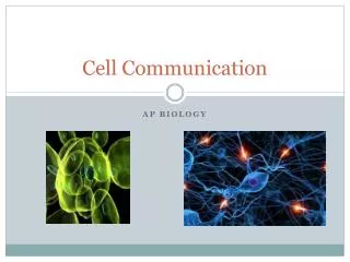
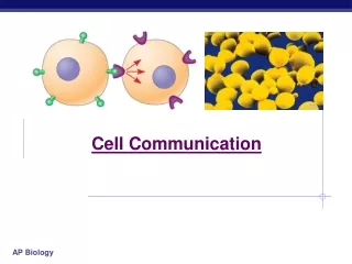
Cell Communication 3 stages of cell communication G Protein-coupled receptors Receptor Kinases Cell signal amplification by phosphorylation Cell response in nucleus (genes) vs. cytoplasm (proteins) Apoptosis
Big Questions: • Why do cells communicate? • What does cellular communication look like? • How is cellular communication utilized in unicellular and multicellular life?
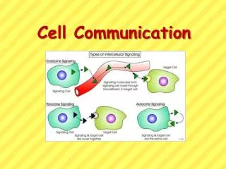
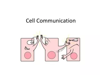
How Cells Communicate: Signal Transduction External signals are converted to responses within the cell. Cells communicate by: • Direct Contact • Short or long distance – by secreting local regulators (growth factors, neurotransmitters)
Direct Contact Plasmodesmata Junctions – Plant and animal cells have junctions that connect the cytoplasm of 2 cells, allowing transfer of materials. Cell-cell recognition – two cells can communicate by interaction with molecules on surfaces
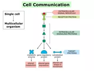
3 Phases of Signal Transduction 1. Reception – The target cell’s detection of a signal molecule coming from outside the cell. 2. Transduction – The conversion of the signal to a form that can bring about a specific response. 3. Response – The specific cellular response to the signal molecule.
How Signals are Received A signaling molecule binds to a receptor protein, causing it to change shape. • Specific fit between signal molecule (LIGAND) and a receptor • Receptors can be INTRACELLULAR or in the PLASMA MEMBRANE.
Intracellular Receptors • Found inside the cell (cytoplasm or nucleus) • Signal molecule must cross plasma membrane (is hydrophobic or very small) Plasma Membrane Receptors • In plasma membrane • Bind to water-soluble ligands
Signaling Pathways I. G Protein Linked Reception Membrane receptor that works with help of a G protein. (G proteins: proteins activated by the transfer of a phosphate from a molecule of GTP.) • Step 1: Ligand binds to G protein – coupled receptor. • Step 2: Conformational change occurs in receptor so it can bind with an inactive G protein • Step 3: a GTP molecule displaces the GDP, activating the G protein • Step 4: G protein binds to a specific enzyme and activates it. • Step 5: Enzyme triggers the next step in the pathway leading to cellular response.
Signaling Pathways II. Receptor Tyrosine Kinases Tyrosine Kinase: Membrane proteins that form dimers (Kinase: enzyme that phosphorylates; Dimer: molecule formed by 2 subunits; Phosphatase: protein that dephosphorylates) • Step 1: Ligands bind to receptors and they form a dimer (Each tyrosine kinase adds a phosphate from an ATP molecule to activate) • Step 2: Receptor protein is activated and starts cellular responses for each phosphorylated tyrosine. Receptor Tyrosine kinases can activate multiple cellular responses (G proteins can only do one).
Receptor Tyrosine Kinase • Receptor Tyrosine Kinase Animation
Signaling Pathways III. Ligand-Gated Ion Channels Specific signal molecules cause ligand-gated ion channels in a membrane to open or close. • Incoming ions trigger the response. • Regulate the flow of specific ions (Facilitated Diffusion)
How Signals are TransducedCascades of molecular interactions relay signals from receptors to target molecules in the cell. • Phosphorylation Cascade – Protein kinases phosphorylate (thus activate) many proteins at the next step. This AMPLIFIES the signal. What kind of feedback is this?
Protein Phosphatases – Enzymes that remove phosphate groups and inactivate kinases. Thus, Signals can be turned ON by kinases and OFF by phosphatases.
Second messengers – Small, non-protein water soluble molecules or ions along a signal transduction pathway that can start a phosphorylation cascade Examples: Calcium Ions Cyclic AMP (affects metabolism)
How Cells Respond Cell signaling leads to regulation of transcription (RNA from DNA) or cytoplasmic activities. • Regulation of protein synthesis by turning specific genes on or off in the nucleus. • Regulation of protein activities in the cytoplasm (ex: activity of enzymes or cytoskeleton rearrangement).
Example: Epinephrine • Aka “Adrenaline” • A common hormone in vertebrates involved in short term stress (“fight or flight” response) • A polar amine ligand • Epinephrine signal transduction is mediated by G-protein linked receptors • It has multiple effects; One response is the inhibition of glycogen synthesis and the acceleration of glycogen break down (Why?)
Complication • A “branching” network • Ex: Wiskott Aldrich syndrome: missing a relay protein interferes with the cytoskeleton • Things get complicated quickly • Simplicity leads to complexity
Why Cells Communicate Examples (Unicellular) • Quorum Sensing – communication among microbes that triggers group response once particular population densities are reached a. Vibrio fischeri– a bacterium that lives inside organs in marine animals. When population density hits a threshold, they begin to produce a light-producing protein. This gives the host animal bioluminescence.
b. Biofilms – bacterial ecosystems that are established and maintained due to elaborate quorum sensing networks. • Plaque Biofilm all on your teeth! • Fruiting body formation in Soil Bacteria in response to poor environmental conditions (How can this kind of behavior evolve?)
II. Yeast Mating – Mating type in (haploid) yeast is genetically determined. • Two mating types: a and alpha. • Each makes signaling molecules that the other receives. • The reception of a mating factor leads to the production of a mating “Shmoo” • Fusion of “schmoos” = diploid yeast cell • Meiosis soon begins
Apoptosis: Cell Suicide Apoptosis integrates multiple cell-signaling pathways. • Elaborate example of cell signaling where a cell is systematically dismantled and digested. • Protects neighboring cells from damage. • In vertebrates, apoptosis is a normal part of the nervous system, immune system, and proper development of hands and feet in humans. Cancer is often a result of disabled apoptosis. Animation.