Download
1 / 67
670 likes | 791 Vues
This comprehensive guide delves into the respiratory system, covering its organs, functions, and the process of respiration. Learn about the upper and lower respiratory tracts, conducting and respiratory portions, along with the vital role of mucous in the respiratory tract. Discover the importance of lungs, bronchial tree, alveoli, and more. Dive into the mechanics of breathing, gas exchange, and pressures involved in respiration. Gain insights into normal inspiration and expiration processes, as well as the differences between costal and diaphragmatic breathing. Enhance your understanding of this crucial system with detailed explanations and visuals.

E N D
Review slides Lecture Exam 2
Respiratory System Respiration (in the respiratory system) is the process of exchanging gases between the atmosphere and body cells. It consists of the following events (in order): • *pulmonary ventilation • *external respiration • transport • internal respiration • cellular respiration Functions of the respiratory system You should know the order of these events. We breathe: 1. To provide O2 for cellular respiration and 2. To rid our bodies of CO2 (waste gas)
Organs of the Respiratory System Upper respiratory tract – nose, nasal cavity, sinuses, and pharynx Lower respiratory tract – larynx, trachea, bronchial tree, lungs Conducting portioncarries air; nose to the terminal bronchioles Respiratory portion exchanges gases; respiratory bronchioles and alveoli
Mucous in Respiratory Tract Respiratory mucosa lines the conducting passageways and is responsible for filtering, warming, and humidifying air. Pseudostratified, ciliated columnar epithelium with goblet cells Respiratory epithelium is interrupted by stratified squamous epithelium in the oro- and laryngopharynx
Nose and Paranasal Sinuses The nose: 1) warms 2) cleans 3) humidifies air Paranasal sinuses are mucus membrane-lined, air-filled spaces in maxillary, frontal, ethmoid, and sphenoid bones that drain into the nasal cavity Figure from: Martini, Anatomy & Physiology, Prentice Hall, 2001 • Sinuses: • Reduce skull weight • Serve as resonating chambers
Inelastic Vestibular folds Covered by folds of laryngeal epithelium that project into glottis Protective Sound Vocal folds (cords) Elastic Larynx Prevents swallowed material from passing into trachea = major components of larynx Posterior Figure from: Martini, Anatomy & Physiology, Prentice Hall, 2001
Trachea & Primary Bronchi Posterior (Smooth muscle) Note that the trachea is anterior to the esophagus (T5) (T6) Anterior C-rings of cartilage: 16-20 incomplete rings completed posteriorly by trachealis musclekeep trachea open (patent) Figures from: Martini, Anatomy & Physiology, Prentice Hall, 2001
Bronchial Tree (No cartilage,few/no smooth muscle) (Cartilage + smooth muscle) (Smooth muscle) Bronchi Bronchioles Alveolar structures Primary Secondary (lobar) Tertiary (segmental) Alveolar ducts Alveolar sacs Alveoli Intralobular Terminal Respiratory Trachea respiratory portion conducting portion Know this chart
Bronchial Tree Hilus of lung is the medial opening for air passageways, blood vessels, nerves, and lymphatics. Carina Bronchi - Primary; w/ blood vessels - Secondary (lobar); two on left, three on right - Tertiary (segmental); supplies a broncho- pulmonary segment; 10 on right, 8 on left Bronchioles - Intralobular; supply lobules, the basic unit of lung - Terminal; 50-80 per lobule - Respiratory; a few air sacs budding from theses Bronchioles are to the respiratory system what arterioles are to the circulatory system Intralobular Figure from: Martini, Anatomy & Physiology, Prentice Hall, 2001
Lobules of the Lung The Lobule is the basic unit of structure and function in the lung (Intralobular) Terminal and respiratory bronchioles are lined with cuboidal epithelium, few cilia, and no goblet cells Figure from: Martini, Anatomy & Physiology, Prentice Hall, 2001
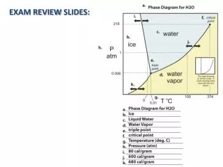
Gases and Pressure • Our atmosphere is composed of several gases and exerts pressure • 78% N2, 21% O2, 0.4% H2O, 0.04% CO2 • 760 mm Hg, 1 ATM, 29.92” Hg, 15 lbs/in2,1034 cm H2O • Each gas within the atmosphere exerts a pressure of its own (partial) pressure, according to its concentration in the mixture (Dalton’s Law) • Example: Atmosphere is 21% O2, so O2 exerts a partial pressure of 760 mm Hg. x .21 = 160 mm Hg. • Partial pressure of O2 is designated as PO2
Normal Inspiration An active process • Intra-alveolar (intrapulmonary) pressure decreases to about 758mm Hg as the thoracic cavity enlarges (P 1/V) • Atmospheric pressure (now higher than that in lungs) forces air into the airways • Compliance – ease with which lungs can expand Phrenic nerves of the cervical plexus stimulatediaphragm to contract and move downward and external (inspiratory) intercostal muscles contract, expanding the thoracic cavity and reducing intrapulmonary pressure. Attachment of parietal pleura to thoracic wall pulls visceral pleura, and lungs follow.
Maximal (Forced) Inspiration Thorax during normal inspiration • Thorax during maximal inspiration • aided bycontraction ofsternocleidomastoid and pectoralis minor muscles Compliance decreases as lung volume increases Costal (shallow) breathing vs. diaphragmatic (deep) breathing
Normal Expiration • due to elastic recoil of the lung tissues and abdominal organs • a PASSIVE process(no muscle contraction involved, no energy needed) Normal expiration is caused by - elastic recoil of the lungs (elastic rebound) and abdominal organs - surface tension between walls of alveoli (what keeps them from collapsing completely?)
Maximal (Forced) Expiration • contraction of abdominal wall muscles • contraction of posterior (expiratory) internal intercostal muscles • An active, NOT passive, process
Terms Describing Respiratory Rate • Eupnea – quiet (resting) breathing • Apnea – suspension of breathing • Hyperpnea – forced/deep breathing • Dyspnea – difficult/labored breathing • Tachypnea – rapid breathing • Bradypnea – slow breathing Know these
Alveoli and Respiratory Membrane • Respiratory Membrane consists of the walls of the alveolus and the capillary, and the shared basement membrane between them Mechanisms that prevent alveoli from filling with fluid: • cells of alveolar wall are tightly joined together • 2) the relatively high osmotic pressure of the interstitial fluid draws water out of them • 3) there is low pressure in the pulmonary circuit Surfactant resists the tendency of alveoli to collapse on themselves.
Diffusion Through Respiratory Membrane The driving for the exchange of gases between alveolar air and capillary bloodis the difference in partial pressure between the gases. alveolus tissues Because O2 and CO2 are relatively insoluble in H2O (plasma), RBCs are used to carry or transform these gases.
Oxygen Transport • Most oxygen binds to hemoglobin to formoxyhemoglobin (HbO2) • Oxyhemoglobin releases oxygen in the regions of body cells • Much oxygen is still bound to hemoglobin in the venous blood Tissues Lungs But what special properties of the Hb molecule allow it to reversibly bind O2?
The O2-Hb Dissociation Curve Recall that Hb can bind up to 4 molecules of O2 = 100% saturation At 75% saturation, Hb binds 3 molecules of O2 on average Sigmoidal (S) shape of curve indicates that the binding of one O2 makes it easier to bind the next O2 This curve tells us what the percent saturation of Hb will be at various partial pressures of O2
Oxygen Release • Amount ofoxygen released from oxyhemoglobinincreases as • partial pressure of carbon dioxide increases • the blood pH decreases and [H+] increases (Bohr Effect; shown below) • blood temperature increases (not shown) • concentration of 2,3 bisphosphoglycerate (BPG) increases (not shown)
Carbon Dioxide Transport in Tissues • dissolved in plasma (7%) • combined with hemoglobin as carbaminohemoglobin(15-25%) • in the form of bicarbonate ions (68-78%) CO2 + H2O ↔ H2CO3 H2CO3 ↔ H+ + HCO3- CO2 exchange in TISSUES
Carbon Dioxide Transport in Lungs CO2 exchange in LUNGS
Summary of Gas Transport PO2 = 40 mm Hg PO2 = 100 mm Hg PO2 = 100 mm Hg PO2 = 40 mm Hg T I S SUES LUNGS PCO2 = 45 mm Hg PCO2 = 40 mm Hg PCO2 = 40 mm Hg PCO2 = 45 mm Hg CO2 + H2O ← H2CO3 ← H+ + HCO3- H+ + HCO3- ← H2CO3 ← CO2 + H2O
Control of Respiration • Control of respiration is accomplished by: 1) Local regulation 2) Nervous system regulation • 1) Local regulation • alveolar ventilation (O2), Blood flow to alveoli • alveolar ventilation (O2), Blood flow to alveoli • alveolar CO2, bronchodilation • alveolar CO2, bronchoconstriction
Control of Respiration • 2) Nervous System Control • The DRG and VRG in medullary respiratory rhythmicity center control rate/depth of breathing • Changes in breathing • CO2 is most powerful respiratory stimulant • Recall: H2O + CO2↔ H2CO3 ↔ H+ + HCO3- • Peripheral chemoreceptors (aortic/carotid bodies) • PCO2, pH , PO2 stimulate breathing • Central chemoreceptors (medulla) • PCO2, pH stimulate breathing
Major Organs of Digestive System Digestion is the mechanical and chemical breakdown of food into a small enough form that cells can absorb • Organs can be divided into the: • Digestive tract (primary) (alimentary canal); tube extending from mouth to anus (about 30 ft.); in contact with food • Accessory organs (secondary); teeth, tongue, salivary glands, liver, gallbladder, and pancreas; provide secretions for digestion • Two major movements stimulating digestion: 1) segmentation and 2) peristalsis
Alimentary Canal Wall Know the 4 layers of the alimentary canal Figure from: Martini, Anatomy & Physiology, Prentice Hall, 2001
Innervation of the Alimentary Canal The alimentary canal has extensive sympathetic and parasympathetic innervation - mainly in the muscularis externa - regulates its tone and the strength, rate, and velocity of muscular contractions • submucosal plexus – controls secretions/blood flow • myenteric plexus – controls gastrointestinal motility/sphincters • parasympathetic division of ANS – increases activities of digestive system and relaxes sphincters • sympathetic division of ANS – generally inhibits digestive actions and contracts sphincters
Palate • roof of oral cavity Figure from: Hole’s Human A&P, 12th edition, 2010 (adenoids) Important in separating the nasopharynx from the pharynx during swallowing Epiglottis prevents food from entering trachea during swallowing
Secondary (Permanent) Teeth Figure from: Hole’s Human A&P, 12th edition, 2010 Total of 32 secondary (permanent) teeth; total of 20 primary (baby, milk) teeth 1 16 Be able to label this diagram I C Big Molars!!! 32 17 Know the order of these
Pharynx Figure from: Hole’s Human A&P, 12th edition, 2010 Pharynx aids swallowing by grasping food and moving it toward the esophagus.
Three Phases of the Swallowing Reflex Only voluntary phase is the buccal (oral) phase, i.e., the initiation of swallowing, then… • soft palate and uvula raise • hyoid bone and larynx elevate Pharyngeal phase • epiglottis closes off top of trachea • longitudinal muscles of pharynx contract reflexive • inferior constrictor muscles relax and esophagus opens Esophageal phase • peristaltic waves push food through pharynx Esophagus conveys food from pharynx to stomach by peristalsis
Stomach Review • Stomach (know all these) • Cardia, fundus, body, pylorus • Mixes food and begins digestion of protein • Limited absorption (alcohol) • Moves food into small intestine • Pyloric sphincter (entrance to small intestine); opens when liquified stomach contents (chyme) exerts enough pressure • Rugae (flatten as it fills) and gastric pits -> gastric juice • Gastric glands • Mucous cells (goblet) – secrete mucous • Chief cells (peptic) – secrete pepsinogen • Parietal cells (oxyntic) – secrete HCl (Parietal, pH); Intrinsic factor for absorption of vitamin B12 • G cells -> gastrin (The Go hormone!); D cells -> Somatostatin (The Stop hormone!)
Three Phases of Stomach Control • Cephalic phase • triggered by smell, taste, sight, or thought of food • begin secretion and digestion • Gastric phase • triggered by distension, presence of food, and rise in pH in stomach • enhance secretion and digestion • Intestinal phase • triggered by distension of small intestine and pH change • controls rate of gastric emptying; may slow emptying; the more fat in the chyme, the slower the emptying NOTE that all the phases control activity in the STOMACH Know what each phase does (shown in red)
Parasympathetic NS G cells Gastrin + Both + Key Overview of Gastric Control/Secretion + Stimulation - Mucous Cells Inhibition Emptying of Stomach ( [H+ ]) Stomach Molility (Segmentation/Peristalsis) ECL Cells Histamine Endocrine Factor + + + Exocrine Factor + (cephalic/gastric phases) + D cells Somatostatin Intrinsic Factor + B12 Parietal Cells pH < 3.0 - + H+ + Cl- HCO3- (alkaline tide) + + + + + (intestinal phase) Stretch of stomach Fats in Small Intestine pH > 3.0(dilution of H+) Peptides Chief Cells Pepsinogen Pepsin Protein Breakdown Food in Stomach Ileum Fat Breakdown Lipases
Pancreatic Juice • pancreatic amylase – splits glycogen into disaccharides • pancreatic lipases – break down triglycerides • pancreaticnucleases – digest nucleic acids • bicarbonate ions – make pancreatic juice alkaline (pH = 8) and neutralize acid (chyme) coming from stomach • Pancreatic proteolytic enzymes…
Pancreatic Proteolytic Enzymes Enteropeptidase (Enterokinase)(brush border of sm. intestine) Know this chart Trypsinogen Trypsin Chymotrypsinogen Chymotrypsin Pancreas Procarboxypeptidase Carboxypeptidase Proelastase Elastase (Proenzymes, Zymogens) (Active enzymes) Dipeptides, tripeptides, amino acids Proteins Purpose of proteolytic enzymes is to continue the breakdown of proteins that began in the stomach
Key Regulation of Pancreas/Intestinal Digestion + Stimulation Acidic Chyme Enters Duodenum + (brush border) + + Enterokinase Cholecystokinin(CCK) Secretin + Trypsinogen Trypsin + + + Gallbladder Contraction Relaxation of hepatopancreatic sphincter ChymotrypsinogenProcarboxypeptidaseProelastase ChymotrypsinCarboxypeptidaseElastase Pancreas Bile and Pancreatic ducts (proenzymes, zymogens) Proteins Bile Lipases HCO3-, PO43- (emulsification) Nucleases(DNA, RNA) Amylase(glycogen, starches) Di- and tripeptides TriglyceridesCholesterolFat Soluble Vitamins pH to ≈ 8 (req. for enzyme action) Nucleotides Mono-, di-, trisaccharides Action of brush border enzymes Fatty acids,monoglycerides Lacteals Portal Vein Amino acids Conversion to chylomicrons Subclavian vein Monosaccharides
Liver Functions (over 200!) • Three general categories of function 1) Metabolic regulation • Interconversion of carbohydrates, lipids, amino acids • Removal of wastes • Vitamin and mineral metabolism • Drug inactivation • Storage of fats, glycogen, iron, vit A/B12/D/E/K 2) Hematological regulation • Phagocytosis and antigen presentation; ab removal • Synthesis of plasma proteins • Removal of circulating hormones • Removal of worn-out RBCs (Kupffer cells) • Removal or storage of toxins 3) Synthesis and secretion of bile (digestion) Know items in red
Paths of Blood and Bile in Hepatic Lobule Figure from: Hole’s Human A&P, 12th edition, 2010 Liver’s role in digestion is production of bile Sinusoid Hepatic portal vein → sinusoids → central vein → hepatic veins → inferior vena cava Hepatic artery
Composition of Bile (Chole-) Yellowish-green liquid continually secreted by hepatocytes • water • bile salts (bile acids) • derived from cholesterol • emulsification of fats (increases surface area for digestive enzymes; large fat blobs become smaller blobs) • absorption of fatty acids, cholesterol, and fat-soluble vitamins • 80% are recycled (reabsorbed and reused) – enterohepatic circulation of bile • 20% excreted in feces (disposes of excess cholesterol) • bile pigments (bilirubin and biliverdin from breakdown of RBCs) • electrolytes The hormone secretin, released by the small intestine, stimulates the hepatocytes to produce a bicarbonate-rich bile that neutralizes acidic chyme coming from the stomach
Gallbladder [Cyst(o)-] Figure from: Martini, Anatomy & Physiology, Prentice Hall, 2001 Main function is to store and concentrate bile between meals, and release concentrated bile under the influence of CCK
Regulation of Bile Release from GB Figure from: Hole’s Human A&P, 12th edition, 2010 • fatty chyme entering duodenum stimulates the GB to release bile (via CCK) Secretin causes the bile ducts (and pancreatic ducts) to secrete bile rich in HCO3-
Actions of Cholecystokinin (CCK) on Digestion Figure adapted from: Barrett, K., Gastrointestinal Physiology, Lange, 2006 CCK Contraction of Gallbladder Secretion of pancreatic enzymes Reduced emptying of stomach Relaxation of hepatopancreatic sphincter Protein, CHO, lipid absorption and digestion Matching of nutrient delivery to digestive and absorptive capability
Small Intestine • Small Intestine • Three major parts • Duodenum – mixing chamber; mucus, buffers, enzymes • Jejunum – digestion and absorption • Ileum – connects to cecum of large intestine • Blood supply and drainage via superior mesenteric artery/vein • Surface area greatly increased, especially in the jejunum, by • Plicae • Villi • Microvilli
Small Intestine (cont’d) • Secretions • mucus secretion (protective) stimulated by presence of chyme in small intestine • distension of intestinal wall activates nerve plexuses in wall of small intestine • motility/secretion stimulated by gastroenteric reflex • parasympathetics trigger release of intestinal enzymes • Absorption • Protein, CHO, electrolytes –> to hepatic portal vein into liver • Fats via chylomicrons and lacteals -> circulation (2nd pass) • Movements • Local via myenteric plexuses • Long distance via stomach filling • Gastroenteric reflex • Gastroileal reflex Know these things…
Secretions of Small Intestine • peptidase – breaks down peptides into amino acids • sucrase, maltase, lactase – break down disaccharides into monosaccharides • intestinal lipase – breaks down fats into fatty acids and glycerol • enterokinase – converts trypsinogen to trypsin • gastrin/somatostatin – hormones that stimulate/inhibit acid secretion by stomach • cholecystokinin (CCK) – hormone that inhibits gastric glands, stimulates pancreas to release enzymes in pancreatic juice, stimulates gallbladder to release bile, and relaxes hepatopancreatic sphincter (of Oddi) • secretin – stimulates pancreas to release bicarbonate ions in pancreatic juice; stimulates gall bladder to release bicarbonate-rich bile Brush border See Table 17.9 in Hole for summary of digestive enzymes
Absorption of Fats in the Small Intestine Figure from: Hole’s Human A&P, 12th edition, 2010 • fatty acids and glycerol • several steps • absorbed into lymph into blood Chylomicrons contain TG, cholesterol, and phospholipids
Functions of Large Intestine • little or no digestive function • absorbs water, bile salts, and electrolytes • secretes mucus (lubrication, binding, protection, pH) • conversion of bilirubin (uro- and stercobilinogen) • houses intestinal flora (~800 species of bacteria) and absorbs vitamins liberated by bacterial action (K, B5, and Biotin); produces intestinal gas (flatus) • forms and stores feces • carries out defecation