Nanowire Biosensors
50 likes | 482 Vues
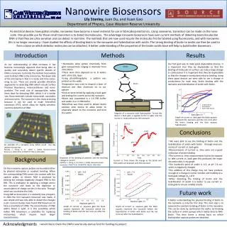
Avidin. O. N. N. Biotin. S. Nanowire. SiO 2. Si Wafer. Lily Stanley, Juan Du, and Xuan Gao Department of Physics, Case Western Reserve University .

Nanowire Biosensors
E N D
Presentation Transcript
Avidin O N N Biotin S Nanowire SiO2 Si Wafer Lily Stanley, Juan Du, and XuanGao Department of Physics, Case Western Reserve University As electrical devices have gotten smaller, nanowires have become a novel material for use in fabricating electronics. Using nanowires, transistors can be made on the nano-scale. One possible use for these small transistors is to detect biomolecules. The advantage nanowire biosensors have over current methods of detecting biomolecules like DNA is that they are ultra-sensitive and can detect in real time. The methods that are now used require the molecules first be labeled using fluorescents, and with nanowires this is no longer necessary. I have studied the effects of binding biotin to the nanowire and hybridization with avidin. The strong binding of biotin to avidin can then be used to form a base on which detector molecules can be attached. A better understanding of the properties of the biotin-avidin base will help to build better biosensors. Introduction Methods Results • Nanowires were grown chemically from gold nanoparticles ranging in diameter from 20 to 40nm • These were then deposed on to Si wafers with a thin SiO2 layer. • Using photolithography a pattern was printed on the wafer • Evaporation was used to deposit a layer of titanium and then aluminum on to our pattern • Devices were tested by applying a back gate and testing the current across the nanowire • Biotin was suspended in a 5.6 PBS buffer and avidin in a 7.4 PBS buffer • Microflow was then used to deliver biotin solution onto device to allow biotin to physically attach to the nanowire and bind the avidin. Our first goal was to make good depleatable devices. It is important that they be depleatable so that the binding of molecules on surface can induce large change in conductance It is important that they be depleatable so that the change in conductance due to binding. Using these good devices we were able to see a change in conductance for each step, biotin binding with the nanowire and hybridization of avidin with biotin. As our understanding of DNA increases it has become increasingly apparent that being able to quickly and accurately detect specific strands of DNA is necessary. Currently the method most widely used to detect DNA is the microarray. The down side of the microarray is that it is costly and time consu-ming to run. There are several possible alterative approaches to detecting DNA which include Surface Plasmon Resonance, microcantilevers and nano-particles. The small size of nanoparticles makes them ideal for detecting DNA, which is of a similar size. Nano-meterials include nanocrystal, nanotubes and nano-wires. The nanowire is the most promsing because it can be used to make field-effect transistors (FET), which allow for highly sensitive, real time detection of DNA. Nanowire Biosensors Diagram of a nanowire with hybridization. To test this device a back gate is applied to the Si wafer and the current is measured across the nanowire Graph of current vs. back gate the black squares represent the nanowire and the red circle after the biotin binding and the blue squares hybridization Conclusions • We were able to see the binding of biotin and the hybridization of avidin with biotin through measure-ments of current vs. back gate • Measurements of current vs. time were not a good indication of protein biding • The current vs. time measurements had to be stopped to take current vs. back gate this produced the major discontinuities in my graph • The isoelectric point of avidin is 6.3, at pH 7.4 our avidin is negatively charged • The addition of this charge may not have produce enough of a change in carrier number and mobility at a back gate voltage (Vg ) of 0 • When detecting the binding of biotin and the hybridization of avidin it is better to use current vs. back gate to ensure reliable results • An example of a nanowire array which could one day replace microarrays • Patolsky, F., Zheng, G., & Lieber, C. (2006). Nanowire-based biosensors. ANALYTICAL CHEMISTRY, 78(13), 4260-4269. Background • Depiction of the binding of proteins and the resulting change in current • Patolsky, F., Zheng, G., & Lieber, C. (2006). Nanowire-based biosensors. ANALYTICAL CHEMISTRY, 78(13), 4260-4269. Current vs. Time shows the change as the biotin and then avidin are deposed onto the nanowire On the nanowire capture probes are bounded either by physical adsorption or covalent bonding. When the corresponding DNA comes into contact with the capture probe, an electric field is produced by binding the strongly negatively charged DNA to the NW. This is analogous to applying a voltage across the nanowire and leads to the depletion or accumulation of charge carriers in the wire. Through which we can detect the DNA. Using NW as biosensors is a relatively new prospect. In 2001 the first nanowire biosensor was made, it was simple and was only able to detect the changes in pH. Current studies have found NW biosensors to have a sensitivity of 10fM. The concentration of DNA within blood is 5μM, it is this sensitivity which makes NW biosensors a candidate for replacing microarrays, which require much larger concentrations. Future work A better understanding the physical binding of biotin to the nanowire is only the first step. The next step is to examine the covalent bonding of biotin to the nanowire. This can be done by sanitization the wafer, terminating the surface in amine groups which reality bind to NHS-biotin. This then forms a strong basis on which biotinylation capture probes are attached. Graph of current vs. aqueous gate the black squares represent the nanowire before the binding of biotin and the red circle are after the binding Graph of current vs. aqueous gate the black squares represent the nanowire before the hybridization of avidin with biotin and the red circle are after the hybridization Acknowledgments I would like to thank the CWRU new faculty startup fund for funding my project.