Download
1 / 29
320 likes | 606 Vues
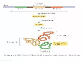
DNA that contains functional information for the synthesis of RNA or Protein → gene Ribosomal RNA ’ s ( rRNA ’ s )→ complex that synthesizes proteins Messenger RNAs ( mRNA ’ s ) → intermediaries → carrier of genetic Info. from 1 or may genes to the ribosome

E N D
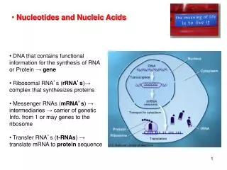
DNA that contains functional information for the synthesis of RNA or Protein → gene • Ribosomal RNA’s (rRNA’s)→ complex that synthesizes proteins • Messenger RNAs (mRNA’s) → intermediaries → carrier of genetic Info. from 1 or may genes to the ribosome • Transfer RNA’s (t-RNAs) → translate mRNA to protein sequence • Nucleotides and Nucleic Acids
Nucleotides have 3 features: • Nitrogen containing bases • Pentose (5 C- sugar) • Phosphate moiety Ester bond Molecule WITHOUT PO4→ Nucleoside NB: ‘ (primers) give to distinguish from C in purine/pyrimidines
Pentose + Purine/Pyrimidine O-glycosidic bond (Nucleoside) + phosphate Nucleotide • Purine and Pyrimidine Bases Proton extraction Break double bond N-b-glycosyl bond with the 1’ C in the pentose (previous slide)
Purines • Present in BOTH DNA and RNA (G) (A)
Pyrimidines • Cytosine (C) found in BOTH DNA and RNA • Thymine (T) found ONLY in DNA • Uracil (U) found ONLY in RNA Distinguishing features A, T G and C nucleotides → genetic information
Two kinds of Pentoses present…… H Deoxy→ deoxyribo(nucleotides/sides) → ribo(nucleotides/sides)
Phosphodiester Bonds link Successive Nucleotides in Nucleic acids • DNA and RNA nucleotides linked via PO4- grp 5’→ 3’-OH grp of the next nucleotide (phosphodiester linkage) • Backbone of alternative linkages b/w PO4 and pentose grps • Backbone is hydrophilic → -OH in CHO/carbohydrate undertake H-H bonding with H2O • PO4 gp pKa = 0, pH 7 → -ve charged → interactions with +ve charges (proteins/metals/polyamines) [Expt. - Adding salt interfers with these interactions = ability to separate DNA from other cellular constituents] • ALLphosphodiester linkage have the SAME orientation 5’→ 3’ (polarity) • 5’ end → LACKS a nucleotide at position 5’ • 3’ end →LACKS a nucleotide at position 3’
DNA/RNA Slow/non-enzymatic hydrolysis Breaking phosphodiester bonds • RNA is more unstable → rapid breakdown in base • (via the 2’-OH) • DNA does NOT • (2’-OH gp is absent)
Nomenclature of nucleotides – what’s in a name… Written in the 5’→ 3’ direction (left to right) Maybe written as: pA-C-G-T-AOH pApCpGpTpA pACGTA Oligonucleotides (<50 nucleotides; >50 – polynucleotides)
DNA is “defined” • DNA base composition varies at a inter-special level • Same species, different tissues have the same DNA base composition • DNA base composition does not change • ALL DNA follows these simple rules: • A = T • G = C • Sum of purines = Sum of pyrimidines: A + G = T + C (Chargeff’s rules) • DNA Structure The famous double helix – solved by x-ray diffraction; insight that molecule was helical; 2 periodicities 3.4Å and 34Å A = T G = C Solve the structure?
Watson and Crick – the master the minds of the jigsaw puzzle 2 helical DNA chain wound around an axis, forming a right handed double helix Hydrophobic backbone of alternative deoxyribose and PO4 gr on outside Pyrimidines/purines stacked inside the double helix (hydrophobic and planar ring structure perpendicular to long axis Offset pairing of 2 stands causes major and minor groove Each paired nucleotide shares the same plane 3 H-H bonds b/w G/C ( G≡C) ;2 b/w A/T (A=T) DNA with high G≡C content difficult to denature 10 base pairs/turn* *crystal form
Complementary strands; two anti-parallel strands containing not identical base bases, yet base pair which fit the A=T and G≡C rules of engagement. Replication
The Chemical Properties of DNA • Highly viscous at Room Temp., pH 7.0 • pH or Temp. (>80oC) → in viscosity Reflect in structural changes Denaturation Melting → disruption of H-H bonding b/w base pairs DS → SS (no covalent bonds are broken) Renaturation is a rapid one step process in undone DS DNA Temp/pH return to physiological conditions → annealing → intact DS In SS DNA, two step process which is slower: “finding” then “zippering” DS – double stranded SS – single stranded
Free nucleotides have the highest UV Absorbance (Abs.) • Once in sequence, stacking of the base pairs ↓ the Abs. (same number/types of nucleotides as above) • Abs. is ↓↓ with complementary pair formation • Denaturation of DS ↑ Abs. HYPERCHROMIC EFFECT • Thus transition from DS to SS can be monitored by UV Abs. Here we can measure the increase and decrease in DNA DS and SS via UV light Some areas more susceptible to denaturation that others…
DNA (viral/Bacterial) denature when heated slowly • Each DNA species has a specific denaturation temp. (Tm) which is reflective of the base pair composition • Higher G≡C content; higher Tm (3 H-H bonds) • A=T bonds require less H to denature • Tm determination can provide estimation of base-pair content • Region with high T=A, denature first (“bubbles”)
Nucleic Acids from different species can form Hybrids The process of complementary sequences in DNA strands can be used to detect similar DNA via duplex formation Denaturing and renealling, complementary regions will bind/seek each other.. Small % will form a mixture of two strands from species – hybrid duplexes→ common evolutionary Heritage, conserved proteins (i.e. conserved DNA/RNA) Gene detection – hybridize ‘seeking’ labeled nucleotide sequence which are site specific. CSI – DNA matching (bone fragments etc.) See changes in UV abs. Thus know if you have a ‘match’ Only small No. will form hybrids
UV light/radiation…..(200-400nm) 2 adjacent Ts Breaking double bond Dimer • Similar effect: • Radiation (C14, H3, X-rays etc.) • Chemicals/alkylating agents • Carcinogens
Sequencing DNA via Sanger Method Separating and identifying DNA strands that differ by a single nucleotide via gel electrophoresis Primer stand has single “non-native” Nucleotides added to it
P OH Add these to our growing sequence BASE (radio labeled nucleotide analogue) H Stops DNA synthesis → a freeze frame of the process of DNA\sequence chain elongation (Dideoxynucleoside)
Analyze the primer at different stages of 5’ chain elongation Labeled Nucleotides Freeze frame Different masses separate MBBE 402 Lecture 15
Nucleotides carry chemical energy in cells Mono-PO4 Di-PO4 Tri-PO4 Sequential hydrolysis Energy
ATP is the most common source for driving energy requiring cellular Rx = 14 KJ/mol Phosphoanhydride bonds = 30 KJ/mol Energy
Adenine Nucleotide are components for many Enzymes cofactors Removal of Adenine → loss of co-factor activity (Vitamin B2)
cAMP, cGMP and ppGpp – used in extracellular chemical signaling • Secondary messengers – target specific membrane bond receptors, which active a cascade of internal cellular events via G-coupled proteins. (bacteria)