Download
1 / 86
860 likes | 1.04k Vues
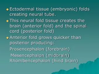
Ectodermal tissue (embryonic) folds creating neural tube. This neural fold tissue creates the brain (anterior fold) and the spinal cord (posterior fold) Anterior fold grows quicker than posterior producing: Prosencephalon (forebrain) Mesencephalon (midbrain) Rhombencephalon (hind brain).

E N D
Ectodermal tissue (embryonic) folds creating neural tube. • This neural fold tissue creates the brain (anterior fold) and the spinal cord (posterior fold) • Anterior fold grows quicker than posterior producing: Prosencephalon (forebrain) Mesencephalon (midbrain) Rhombencephalon (hind brain)
Prosencephalon 1- Telencephalon - Cerebrum (cerebral cortex, white matter and basal nuclei) 2- Diencephalon - (thalamus and hypothalamus)
Mesencephalon - Brain stem (midbrain) • Rhombencephalon 1- Metencephalon -Brain stem (pons) and Cerebellum 2- Myelencephalon - Brain stem (medulla oblongata)
Hollow spaces in the brain termed ventricles. CSF is produced and circulated (lined with ependymal cells) Lateral ventricles (2) separated by a thin membrane called the septum pellucidum. (CSF produced in the choroid plexus) Each lateral ventricle communicates with the third ventricle via a channel called an interventricular foramen (foramen of Monro)
Third ventricle is continuous with the fourth ventricle via the cerebral aquaduct of Sylvius Fourth ventricle drains CSF into spinal cord through the foramen of Magendie.
Cerebral Hemispheres Superior part of the brain • Covered entirely by ridges (gyri), separated by shallow grooves (sulci) and deeper called grooves (fissures) • Anatomical landmarks: • Longitudinal fissure • Frontal, parietal, occipital, and temporal lobes • Deep to temporal, parietal, and frontal lobes is a fifth lobe called the insula • Central sulcus of Rolando • Pre/post central gyri • Lateral sulcus of Sylvius • Calcarine sulcus • Parito-occipital sulcus • Transverse cerebral fissure
Cerebral Cortex: Seat of consciousness. • Cerebral cortex contains three functional areas: 1- Motor areas - control voluntary motor function 2- Sensory areas - provide for conscious awareness of sensation 3- Association areas - integrate all other information Each hemisphere is concerned with the sensory and motor functions of the opposite side of the body
Motor regions are located in the posterior frontal lobe. • Primary motor cortex - precentralgyrus in the frontal lobe - Large neurons (pyramidal cells) allow conscious control of movement of skeletal muscles -The pyramidal cells' long axons from voluntary motor tracts called pyramidal (corticospinal) tracts -Motor areas have been spatially mapped = somatotropy. (Homunculis)
Premotor cortex - anterior to the precentral gyrus in the frontal lobe. Regions controls learned motor skills that are repeated or patterned. Also coordinates the movements of muscles simultaneously and\or sequentially by sending activating impulses to the primary motor cortex.
Broca's area - anterior to the premotor area - Involved in directing motor speech.
Frontal eye field - anterior to the premotor cortex and superior to Broca's area Controls voluntary movement of eyes.
Sensory Areas (parietal, temporal, and occipital lobes) • Primary somatosensory cortex - postcentral gyrus of parietal lobe (immediately behind primary motor cortex) • Neurons receive info (from sensory receptors, skin, and muscles) and identifies body region being stimulated • Somatosensory association area - lies posterior to the primary somatosensory cortex • Integrate and analyze somatic sensory inputs (e.g. temperature and pressure) into comprehensive evaluation.
Visual areas - occipital lobes contain primary visual cortex (receive information from retina) and visual association area (interprets information from retina). • Auditory areas - temporal lobes contain primary auditory cortex (receives impulses from inner ear) and auditory association area (interprets sound).
Olfactory cortex - temporal lobe in region called the uncus; enables conscious awareness of odors. “Skunkus in my uncus” • Gustatory cortex - parietal lobe deep to temporal lobe; involved in perception of taste.
Association Areas • Somatosensory cortex - posterior to the primary somatosensory cortex • The association areas, in turn, communicate with the motor cortex and with other sensory association areas to analyze, recognize, and act on sensory inputs.
Prefrontal cortex - anterior portions of frontal lobe - Involved with intellect and complex learning (cognition) and personality - Tumors may lead to personality disorders - prefrontal lobotomy are performed in severe cases of mental illness. Alzheimer’s Disease
Gnostic area - undefined area in temporal, occipital, and parietal lobes - Receives input from all sensory association areas and stores complex memory patterns associated with sensation - Sends assessment of sensations to prefrontal cortex which adds emotional overtones - Injury to gnostic area causes one to become an imbecile - interpretation to various sensations/stimuli lost.
Language areas - found in Wernicke's area of temporal lobe of one hemisphere (usually left) - Involved in interpretation of language.
Cerebral White Matter: • Responsible for communication within the brain between each hemisphere, cerebral cortex and lower CNS centers. • White matter bundled into large tracts • Fibers and tracts are classified according to the direction in which they run.
Commissural • Connects the gray areas of both hemispheres so the brain functions as one unit • Corpus callosum: superior to the lateral ventricles • Fibers run horizontal Association fibers: - Connect within the same hemisphere. - Connects adjacent gyri and different lobes. - Fibers run horizontal
Projection fibers: • Connects cortex to lower brain centers (spinal cord and brain stem). • Fibers run vertical • Internal capsule and corona radiata
Basal nuclei: (cell bodies) ganglia: Receive extensive input from the entire cerebral cortex and project messages (via relays) to the premotor and prefrontal cortices Structures: Corpus Striatum, composed of • Caudate nucleus • Lentiform nucleus, composed of - Putamen - Globus pallidus Amygdala: hangs off of the tail of the caudate nucleus. Center for fear.
Diencephalon (thalamus, hypothalamus) • Thalamus: • Gray matter areas enclose the third ventricle. • Receive and projects fibers from the cerebral cortex. • All senses (afferent) from the body will pass through the thalamus (relay center). Senses are then sorted out • Gateway to the cerebral cortex
Hypothalamus: • Walls of hypothalamus (tissue) meet and extend, forming infundibulum (stalk), connecting the pituitary to base of hypothalamus. • The hypothalamus is the main visceral control center of the body. • Homeostatic roles: 1- Autonomic control center - regulates involuntary nervous system activity (influences BP, HR, GI motility, Respiration rate and depth, and pupil size).
2- Center for emotional response and behavior - numerous connections with cortical association areas (initiates physical expressions of emotion). 3- Body temperature regulation - hypothalamus neurons monitor blood temperature flowing through hypothalamus (initiates sweating, shivering, etc.). 4- Regulation of food intake - responds to hormones and blood levels of nutrients. (hunger; satiation)
5- Regulation of water balance and thirst - triggers ADH release, causing kidneys to retain water; thirst centers stimulated, cause us to drink. 6- Sleep-wake cycle regulation - set timing of our sleep cycle in response to daylight-darkness cues. 7- Control of endocrine system - hypothalamus produces releasing hormones that control the secretion of anterior pituitary hormones
Brainstem: • Gray matter surrounded by white matter • Controls automatic functions necessary for survival (cardiac, breathing, digestion, head and eye movement) • Composed of the - midbrain - pons - medulla oblongata
Midbrain: Structures: Cerebral peduncles - pyramidal motor tracts descending toward spinal cord. Cerebral aqueduct - connect 3rd and 4th ventricle, enclosed by nuclei Nuclei - corpora quadragemina (two pair) • Superior colliculi (visual reflex, head/eye movement) • Inferior colliculi (auditory relay, startle reflex) Also embedded in white matter of midbrain are two pigmented nuclei Substantia nigra (site of dopamine production) and red nucleus (contains iron & hemoglobin and coordinates muscular movements: limb flexion).
Classes of Neurotransmitters: • Acetylcholine: excitatory (skeletal muscle) • Biogenic amines • Dopamine, Norepinephrine, and epinephrine (feel good catecholamines) • Serotonin: inhibitory • Histamine • Amino acids • GABA (gamma-aminobutyric acid) (inhibitory) • Glutamate: excitatory • Peptides • Endorphins and enkephalins: inhibitory (opioids)