Muscle Quality Assessed Through Ultrasonography Relates to Strength and Function in Older Adults
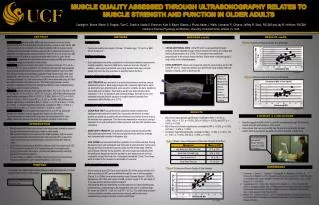
This study investigates the relationship between muscle quality (MQ), assessed via ultrasonography, and muscle strength and function in older adults. Twenty-six healthy participants (13 males and 13 females, average age 71) underwent various tests including hand grip strength, gait speed, chair rise time, and leg-extension strength assessment. Results showed significant correlations between MQ of the vastus lateralis and rectus femoris muscles and measures of strength and function. The findings suggest that improving MQ could enhance daily living activities for older adults.

Muscle Quality Assessed Through Ultrasonography Relates to Strength and Function in Older Adults
E N D
Presentation Transcript
MUSCLE QUALITY ASSESSED THROUGH ULTRASONOGRAPHY RELATES TO MUSCLE STRENGTH AND FUNCTION IN OLDER ADULTS Carleigh H. Boone, Maren S. Fragala, Tyler C. Scanlon, Nadia S. Emerson, Kyle S. Beyer, Gabriel J. Pruna, Adam J. Wells, Leonardo P. Oliveira, Jeffrey R. Stout, FACSM and Jay R. Hoffman, FACSM Institute of Exercise Physiology and Wellness, University of Central Florida, Orlando, FL, USA ABSTRACT METHODS RESULTS cont’d. METHODS cont’d. • Ultrasound measures: • CROSS-SECTIONAL AREA of the RF and VL was quantifiedthrough analysis of three separate images of each muscle from which an average was derived (Koppenhaver, etal.2009). The transducer was positioned perpendicular to the muscle tissue interface. Scans were conducted using LV (logiq view) mode ultrasonography. • ECHO-INTENSITY values were measured using the same images as for CSA of the RF and VL. Grayscale analysis was performed using ImageJ(National Institute of Health, USA) to determine EI. BACKGROUND: Muscle quality (MQ) has been shown to be more closely related to physical functioning than muscle quantity or mass in older adults. MQ can be analyzed non-invasively with ultrasonography (US) to assess muscle architectural characteristics and echo-intensity (EI), a value that measures the reflectivity of intramuscular connective tissue. However, it is not known how MQ analyzed with US relates to muscular strength and function in older adults. PURPOSE: To examine the relationship between ultrasound MQ and measures of muscle strength and function in older adults. METHODS: Twenty-six healthy older adults (13 male, 13 female; 71.0 ± 6.5 y; 27.5 ± 4.8 kg∙m-2) were recruited for this study. Muscle strength was assessed by an individualized bilateral leg-extension multiple-repetition maximum (RM) test. Muscle function was assessed using hand grip dynamometer (HG), gait speed (GS), and chair rise time (CRT). Cross-sectional area (CSA) and EI of the vastus lateralis (VL) and rectus femoris (RF) were measured using US. MQ was determined as CSA relative to EI. The relationship between muscle qualitative variables and muscular strength and function were evaluated using Pearson product moment coefficients. RESULTS: Muscle strength and function were RM = 77.7 ± 31.7 kg, HG = 33.5 ± 12.5 kg, GS = 1.4 ± 0.4 m·s-1, CRT = 13.7 ± 2.6 s. MQ of the vastus lateralis significantly correlated to RM (r = 0.529, p = 0.006), HG (r = 0.721, p = 0.000), GS (r = 0.609, p = 0.003), and CRT (r = -0.416, p = 0.039). MQ of the rectus femoris significantly correlated to RM (r = 0.505, p = 0.010) and HG (r = 0.676, p = 0.000). Combined thigh MQ significantly correlated to RM (r = 0.503, p = 0.010), HG (r = 0.801, p = 0.000), and GS (r = 0.554, p = 0.007). CONCLUSIONS: Results suggest that MQ of the VL and RF as assessed through US relates to muscle strength and function in older adults. Interventions that can improve MQ may have functional benefits for older adults, which may improve the ability to perform activities of daily living. • PARTICIPANTS: • Twenty-six healthy older adults (13 male, 13 female; age: 71.0 ± 6.5 y; BMI: 27.5 ± 4.8 kg∙m-2) • TESTING PROTOCOL: • Each participant voluntarily completed an individualized bilateral leg-extension multiple-repetition maximum (RM) test to measure muscular strength. In addition, each subject performed a hand grip dynamometer test, and gait speed and chair rise time exercises to quantify muscle function. • MUSCLE STRENGTH MEASUREMENT: • LEG STRENGTH was assessed on a seated leg extension machine using a submaximal protocol. Each subject began with a relatively light load to warm up. During the test, all participants were asked to complete as many repetitions as possible prior to fatigue. Their testing weight was determined to allow completion of 5 to 10 repetitions until volitional fatigue. Strength levels were estimated using the Brzycki equation to predict maximal strength from submaximal effort (McNair, et al. 2011). • MUSCULAR FUNCTION MEASUREMENT: • CHAIR RISE TEST was performed to assess functional mobility. Each participant was instructed to stand five consecutive times from a seated position as quickly as possible with arms folded across his/her chest to ensure the exercise was unassisted. Their time was measured in seconds (s) using a stopwatch from each participant’s initial movement until their last repetition was completed. • HAND GRIP STRENGTH was assessed using a hand-grip dynamometer. Each participant performed 2 five-second grip tests from which an average was computed and recorded in kilograms (kg). • GAIT SPEED was examined with the completion of an 8-foot walk test. During the assessment, each participant was instructed to walk at his/her normal pace through an 8-foot course that was previously marked off with tape. With the use of Brower Infrared Timing Systems, the timer began automatically when the participant stepped across the starting line and stopped when one foot completely crossed the finish line. Participants completed 2 trials. Their times were recorded to the nearest one hundredth of a second. • DATA COLLECTION AND ANALYSES: • Cross-sectional area (CSA) and echo-intensity (EI) of the vastus lateralis (VL) and rectus femoris (RF) were quantified through the use of ultrasonography (Figure 1). A 12MHz linear probe scanning head (General Electric LOGIQ P5, Wauwatosa, WI, USA) with a gain of 50dB, dynamic range of 72, and depth of 5 cm was used to optimize spatial resolution. • Individual leg MQ was determined as CSA relative to EI. Ultrasound MQ was determined as combined thigh CSA multiplied by the sum of combined thigh EI, shown as (CSA RF + CSA VL) X (EI RF + EI VL). The relationship between muscle qualitative variables and muscular strength and function were evaluated using Pearson product moment coefficients. Figure 3: Ultrasound Muscle Quality & Leg Strength *p = 0.010 Figure 1: Ultrasound image of the dominant leg Rectus Femoris. Cross-sectional area outlined in yellow. Figure 4: Ultrasound Muscle Quality & Gait Speed *p= 0.007 RESULTS SUMMARY & CONCLUSIONS • MQ of the vastus lateralis significantly correlated to RM (r = 0.529, p = 0.006), HG (r = 0.721, p = 0.000), GS (r = 0.609, p = 0.003), and CRT (r = -0.416, p = 0.039). • MQ of the rectus femoris significantly correlated to RM (r = 0.505, p = 0.010) and HG (r = 0.676, p = 0.000). • Combined thigh MQ significantly correlated to RM (r = 0.503, p = 0.010), HG (r = 0.801, p = 0.000), and GS (r = 0.554, p = 0.007). • Results suggest that MQ of the VL and RF as assessed through US relates to muscle strength and function in older adults. • Interventions that can improve MQ may have functional benefits for older adults, which may improve the ability to perform activities of daily living. INTRODUCTION • Muscle quality (MQ) has been shown to be more closely related to physical functioning than muscle quantity or mass in older adults. • MQ can be analyzed non-invasively with ultrasonography (US) to assess muscle architectural characteristics and echo-intensity (EI), a value that represents the amount of intramuscular connective tissue and adipose tissue. • Echo-intensity values are quantified in arbitrary units of a scale from 0-255. • An increased EI is indicative of increased amounts of connective and adipose tissue within the muscle, and thus decreased levels of strength, power, and functional capacity. • In a prior study, a negative correlation was found between echo-intensity values and strength in older males (Fukumoto, et al. 2012) An ultrasound of a participant’s Rectus Femoris using GE LOGIQ P5 imaging Table 1: Mean values of Measures of Muscular Strength & Function PURPOSE REFERENCES Figure 2: Ultrasound Muscle Quality & Grip Strength • To examine the relationship between ultrasound MQ and measures of muscle strength and function in older adults. Fukumoto, Y., Ikezoe, T., Yamada, Y., Tsukagoshi, R., Nakamura, M., Mori, N., Kimura, M., Ichihashi, N., 2012. Skeletal muscle quality assessed from echo intensity is associated with muscle strength of middle-aged and elderly persons. Eur. J. Appl. Physiol. 112, 1519-1525. Koppenhaver, S.L., Parent, E.C, Teyhen, D.S., Hebert, J.J., Fritz, J.M, 2009. The effect of averaging multiple trials on measurement error during ultrasound imaging of transversusabdominis and lumbar multifidus muscles in individuals with low back pain. J Orthop Sports PhysTher. 604-611. McNair, P.J., Colvin, M., Reid, D., 2011. Predicting Maximal Strength of Quadriceps from Submaximal Performance in Individuals with Knee Joint Osteoarthritis. Am Coll Rheum. 216-222. Bilateral Leg-Extension Multi-Repetition Test *p = 0.000