Download
1 / 19
220 likes | 505 Vues
Lec . 8 Virus genome replication Dr. Ahmed K. Ali. The genome of the infecting virus is replicated so that viral transcription can be amplified and to provide copies of the genome for progeny virions .

E N D
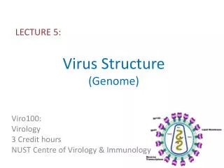
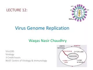
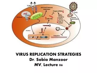
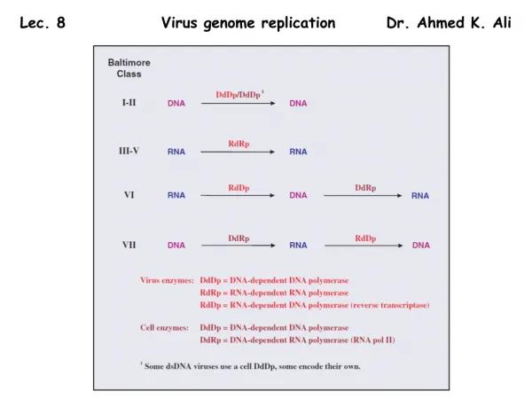
Lec. 8 Virus genome replication Dr. Ahmed K. Ali
The genome of the infecting virus is replicated so that viral transcription can be amplified and to provide copies of the genome for progeny virions. Generally, DNA viruses copy their genomes directly to DNA and RNA viruses copy their genomes directly to RNA. There are, however, some DNA viruses that replicate their genomes via an RNA intermediate and some RNA viruses that replicate their genomes via a DNA intermediate. The various replication modes of virus genomes are summarized in Figure 7.1. Single-stranded genomes are designated as plus or minus depending on their relationship to the virus mRNA. Plus strand genomes have the same sequence as the mRNA (except that in DNA thymine replaces uracil), while minus-strand genomes have the sequence complementary to the mRNA. Single-stranded DNA is converted to dsDNA prior to copying. There are two classes of viruses with (+) RNA genomes (Figure 7.1). Class IV viruses copy their (+) RNA genomes via a (−) RNA intermediate, while Class VI viruses replicate via a DNA intermediate. The synthesis of DNA from an RNA template (reverse transcription) is also a characteristic of Class VII viruses.
Location of virus genome replication in eukaryotic cells The genomes of most DNA viruses are replicated in the nucleus, but those of some dsDNA viruses are replicated in the cytoplasm. The genomes of most RNA viruses are replicated in the cytoplasm, but those of the minus-strand RNA viruses with segmented genomes are replicated in the nucleus. The retroviruses and pararetrovirusesare special cases: each replicates RNA to DNA in the cytoplasm and DNA to RNA in the nucleus.
Initiation of genome replication Each virus genome has a specific sequence where nucleic acid replication is initiated. This sequence is recognized by the proteins that initiate replication. Nucleic acid replication requires priming, which is the first reaction of a nucleotide with an –OH group on a molecule at the initiation site. RNA and protein molecules are required to act as primers to initiate the replication of many DNA and RNA genomes. Cellular DNA synthesis begins after a region of the double helix is unwound by a helicase and after short sequences of RNA (primers) complementary to regions of DNA are synthesized by the action of primase. Some viruses, such as polyomaviruses, use the cell primase to synthesize their RNA primers, while others, such as herpesviruses and phage T7, encode their own primases. For some viruses the primer for initiation of nucleic acid replication is the –OH group on a serine or tyrosine residue in a protein. DNA viruses that use protein primers include some animal viruses (e.g. adenoviruses) and some phages (e.g. tectiviruses). RNA viruses that use protein primers include some animal viruses (e.g. picornaviruses) and some plant viruses (e.g. luteoviruses). Hepadnaviruses are DNA viruses that use a protein primer to initiate (−) DNA synthesis and an RNA primer to initiate (+) DNA synthesis.
Polymerases (Figure 7.3) A DNA virus requires a DNA-dependent DNApolymerase. Amongst the DNA viruses that replicate in the nuclei of eukaryotic cells, viruses with small genomes (e.g. papillomaviruses) use the cell enzyme, while viruses with large genomes (e.g. herpesviruses) encode their own enzyme. Those DNA viruses that replicate in the cytoplasm must encode their own enzyme. The enzyme that replicates the genome of an RNA virus is often referred to as a replicase; for many RNA viruses this is the same enzyme as that used for transcription. The retroviruses and the pararetroviruses encode reverse transcriptasesto transcribe from RNA to DNA, and use the host cell RNA polymerase II to transcribe from DNA to RNA. DNA replication The viruses of Class I (dsDNA) and Class II (ssDNA) replicate their genomes via dsDNA. The ssDNA viruses first synthesize a complementary strand to convert the genome into dsDNA.
Double-stranded RNA replication Double-stranded RNA, like dsDNA, must be unwound with a helicase in order for the molecule to be replicated. Some dsRNA viruses, e.g. Pseudomonas phage ϕ6 (ϕ = Greek letter phi), replicate their genomes by a semi-conservative mechanism, similar to dsDNA replication; each of the double-stranded progeny molecules is made up of a parental strand and a daughter strand. Other dsRNA viruses, including members of the family Reoviridae, replicate by a mechanism designated as conservative because the double-stranded molecule of the infecting genome is conserved (Figure 7.6).
Single-stranded RNA replication The ssRNA genomes of viruses in Classes IV and V are replicated by synthesis of complementary strands of RNA that are then used as templates for synthesis of new copies of the genome (Figure 7.1). The synthesis of each RNA molecule requires the recruitment of an RNA-dependent RNA polymerase to the 3 end of the template, therefore both plus- and minus-strand RNA must have a binding site for the enzyme at the 3 end. Reverse transcription Some RNA viruses replicate their genomes via a DNA intermediate, while some DNA viruses replicate their genomes via an RNA intermediate (Figure 7.1). Both of these modes of genome replication involve reverse transcription, which has two major steps: synthesis of (−) DNA from a (+) RNA template followed by synthesis of a second DNA strand (Figure 7.7). Both steps are catalysed by a reverse transcriptase that is encoded by the virus.
Once threshold quantities of progeny virus genomes and structural proteins have accumulated in the infected cell, assembly of virions can commence. These components are assembled into nucleocapsids. If the virion also contains lipid then the assembly process also includes the acquisition of this component, either as an internal membrane or as an envelope. Nucleocapsid assembly Helical viruses: The assembly of virions and nucleocapsids of ssRNA viruses with helical symmetry involves coating the genome with multiple copies of a protein (Figure 8.1).
Icosahedral viruses: The assembly of virions and nucleocapsids of many viruses with icosahedral symmetry involves the construction of an empty protein shell, known as a procapsid, or a prohead in the case of a tailed bacteriophage. The procapsid is filled with a copy of the virus genome (Figure 8.2); during or after this process it may undergo modification to form the mature capsid. Modification of the procapsidmay result in a change from a spherical to an icosahedral shape. For some viruses, including adenoviruses and picornaviruses, modification of the procapsidinvolves cleavage of one or more of the structural proteins. The genome enters the procapsid through a channel located at a site that will become one of the vertices of the icosahedron. Any enzymes involved in packaging the genome are located at this site.
Genome packaging It has been shown that genome packaging is achieved through the recognition by a virus protein of a specific virus genome sequence, known as a packaging signal. In single-stranded genomes the packaging signal is within a region of secondary structure. Most viruses with single-stranded genomes package either the plus strand or the minus strand, so the packaging signal must be present only in the strand to be packaged. Virus genomes are packaged into small volumes, which means that repulsion between the negative charges on their phosphate groups must be overcome. This may be aided by packaging basic proteins, which are positively charged, along with the genome. Some ssRNAviruses (e.g. rhabdoviruses, influenza viruses and retroviruses) coat their genomes with basic proteins, while some dsDNA viruses (e.g. adenoviruses and baculoviruses) have a basic protein closely associated with the genome.
Assembly mechanisms For some viruses infectious virions can be reassembled from the purified components (protein and nucleic acid), under appropriate conditions of pH and in the presence of certain ions. The viruses that can self-assemble in this way are those with a relatively simple virion composed of a nucleic acid and one or a small number of protein species. Viruses that can self-assemble in a test tube are assumed to undergo self-assembly in the infected cell. Examples are tobacco mosaic virus (helical symmetry) and the ssRNAphages (icosahedral symmetry). Self-assembly is economical because no additional genetic information is needed for the assembly process. The virions of more complex viruses, such as herpesvirusesand the tailed phages, do not reassemble from their components in a test tube. The environment within the infected cell is required and the virionsare constructed by a process of directed assembly. Directed assembly of icosahedral viruses may involve proteins that are temporarily present while the virionis under construction, but are not present in the mature virion. These proteins are known as scaffolding proteins. Once their job is completed they are removed from the procapsid. Some are removed by proteolysis, while others remain intact and can be recycled. Scaffolding proteins of the tailed phages play roles in determining the size and shape of the phage head.
Formation of virion membranes • Budding through cell membranes Most enveloped viruses acquire their envelopes by budding through a membrane of the host cell (Figure 8.3). For viruses with eukaryotic hosts this membrane is often the plasma membrane; the virions of most retroviruses and rhabdoviruses acquire their envelopes in this way. Regions of membrane through which budding will occur become modified by the insertion of one or more species of virus protein, the vast majority of which are glycoproteins. Budding of virions involves interaction between the cytoplasmic tail of a virus glycoprotein in the membrane and another virus protein. In a number of virus groups, including paramyxoviruses and rhabdoviruses, this protein is the M (membrane, matrix) protein (Figure 8.4(a)). M proteins have an affinity for membranes, and bind to nucleocapsids as well as to the virus glycoproteins, ‘stitching’ the two together during budding. Not all enveloped viruses have a layer of protein between the envelope and the nucleocapsid (Figure 8.4(b)). In the virions of alphaviruses (e.g. yellow fever virus) the surface of the nucleocapsid interacts directly with the cytoplasmic tails of the glycoproteins in the membrane.
The late stage of budding involves the membrane pinching off and the release of the newly formed virion.
2. De novo synthesis of viral membranes A minority of viruses direct the synthesis of lipid membrane late in the replication cycle. In some cases the membrane forms a virion envelope (e.g. poxviruses); in other cases the membrane forms a layer below the surface of the capsid (e.g. iridoviruses).
Virion exit from the infected cell The virions of many viruses are released from the infected cell when it bursts (lyses), a process that may be initiated by the virus. Many phages produce enzymes (lysins, such as lysozymes) that break bonds in the peptidoglycan of the host bacterial cell walls. Other phages synthesize proteins that inhibit host enzymes with roles in cell wall synthesis; this leads to weakening of the cell wall and ultimately to lysis. Because of their modes of transmission, most plant viruses leave their host cells in ways that differ from those of animal and bacterial viruses. Plant cells are separated from each other by thick cell walls, but in many of them there are channels, called plasmodesmata, through which the plant transports materials. Viruses are able to spread within the host by passing from cell to cell through plasmodesmata.