Female Breast Physiology: Hormones and Function
Explore the physiological anatomy of the breast, hormonal influences during mammogenesis and lactation, and the phases of lactogenesis. Learn the control mechanisms of lactation and the role of suckling reflex. Dive into the structure of the breast and mammary glands.

Female Breast Physiology: Hormones and Function
E N D
Presentation Transcript
Reproductive Physiology Lecture 8 Hormones affecting female breast Dr.MohammedAlotaibi Assistant Professor dEpartment of Physiology College of Medicine King Saud University
Objectives By the end of this lecture, you should be able to: Know the physiologic anatomy of the breast. Describe the physiological changes that occur in the breast during mammogenesis, lactogenesis, and galactopoeisis and the hormones involved. Recognize the phases of lactogenesis. Describe the endocrine and autocrine control of lactation. Explain the physiological basis of suckling reflex and its role in lactation.
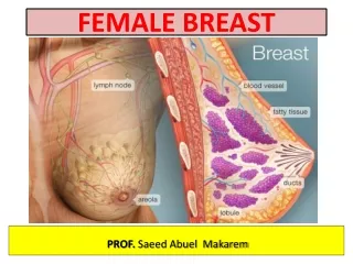
Structures of the Mammary Gland • Each breast consists of ~ 23 lobes of secretorytissue • a. Each lobe has one lactiferous duct • b. Lobes (and ducts) are arranged radially • c. Lobes are composed of lobules • d. Lobules are composed of alveoli
fat nipple Lactiferous sinus Montgomery glands Lactiferous duct areola Alveoli (alveolar gland) Suspensory ligament (Cooper) lobules fat Lobe nipple Interlobular connective tissue
Ductal System • Alveolar tubule • Secondary tubule • Mammary duct • Ampulla (lactiferous sinus) • Lactiferous duct
Lobule-Alveolar System One cell layer The function of the alveolar epithelial cells is to remove nutrients from the blood and transform these nutrients into the components of milk.
Where does milk come from? The fundamental secretory unit of the breast is the alveolus
Stages of Mammary Gland Development 1) Mammogenesis(growth and develpoment of mammary gland to a functional state). 2) Lactogenesis(initiation of milk secretion): Phase 1 Phase 2 3) Galactopoiesis(maintenance of milk secretion in the postpartum period) 4) Involution (cessation of milk production)
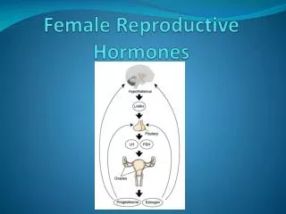
Hormones affecting the female breast • Mammogenic hormones(promoting the proliferation of alveolar and duct cells). • Lactogenic hormones(promoting initiation of milk production by alveolar cells). • Galactokinetichormones (promoting contraction of myoepithelial cells, and thus milk ejection). • Galactopoietic hormones (maintaining milk production afterit has been established).
Mammogenic Hormones (Hormones Involved in Mammary Growth) Estrogens Progesterone Growth hormone Cortisol Insulin (IGF-1) hPL also called hCS Prolactin (PRL) During puberty (ovarian hormones stimulate mammary growth) • During pregnancy (complete development of mammary glands)
Ovarian Hormones • Estrogen • Growth & branching of ductal system (with GH) • Fat deposition in the stroma. • Progesterone • Growth of lobule-alveolar system (budding of alveoli and secretory changes in epithelial cells). Although progesterone and estrogen are essential for physical development of the breasts, they inhibit actual secretion of milk during pregnancy.
Lactogenesis • Lactogenesis: Cellular changes by which mammary epithelial cell switches from non-secretory tissue to a secretory tissue (initiation of milk secretion). • Involves 2 Phases: • Lactogenesis 1 • Lactogenesis 2
Lactogenesis • Lactogenesis 1: (Histological and enzymatic differentiations of mammary epithelial cells). • Starts in mid-pregnancy and characterized by expression of many genes involved in the synthesis of milk components (increases in uptake transport systems for amino acids, glucose, and calcium required for milk synthesis). • Prolactin stimulates mammary secretory cells to produce milk. • Further differentiation is inhibited by high levels of progesterone from the placenta.
Lactogenesis • Lactogenesis 2: (Copious secretion of all milk components), starts 2-3 days postpartum • At parturition, withdrawal of progesterone + high level of prolactin leads to: • Further increase in expression of milk protein genes • Glands absorb large quantities of metabolic substrates from the blood • Movement of cytoplasmic lipid droplets and casein into alveolar lumen • Transfer of immunoglobulins • Secretion of colostrum followed by milk • Suckling stimulates further increase in expression of genes involved in milk secretion with expansion of alveolar epithelium • Lactation is maintained by removal of milk • Switch from endocrine to autocrine control of milk production.
(LactogenicHormones) Prolactin hPL (hCS) Growth hormone Insulin (IGF-1) Cortisol Thyroid hormones parathyroid hormones Withdrawal of estrogens and progesterone All are required to facilitate the mobilization of nutrients and minerals.
Prolactin (PRL) • Secreted from the anterior pituitary gland. • Its level rises steadily from the 5th week of pregnancy until birth (10-20 times the nonpregnant level) (enhanced by E) • It has mammogenic, lactogenic and galactopoietic effects. • It stimulates expression of genes that encode several milk components (casein/ lactalbumin, lactose and lipids) • Sudden drop in E & P after delivery allows milk production • It is controlled mainly by hypothalamic hormone(Dopamine)
Human placental lactogen (human chorionic somatomammotropin, hCS) • Secreted by the placenta at about the 5th week of pregnancy • Its secretion increases progressively throughout the remainder of pregnancy in direct proportion to the weight of the placenta • Causes at least partial development of the animal’s breasts and in some instances causes lactation [facilitates growth of mammary glands, supports prolactin during pregnancy (lactogenic properties)] • It has weak actions similar to those of growth hormone • It decreases maternal insulin sensitivity, decreases utilization of glucose, and promotes the release of free fatty acids (Metabolic)
The alveolar cell secretes the components of milk through five pathways IgA
Galactopoeisis • Galactopoeisis is defined as the maintenance of lactation once lactation has been established. starts 9-15 days postpartum (GalactopoieticHormones) PRL (primary) Cortisol and other metabolic hormones (permissive)
Oxytocin and psychic stimuli initiate milk ejection (“let-down”) • Milk Ejection Reflex: Oxytocincontracts the myoepithelial cells, forcing milk from the alveoli into the ducts and sinuses where it is removed by the infant (galactokinetic effect). • (GalactokineticHormones) Oxytocin (OT) Vasopressin
Autocrine Control of Lactation Influence of Local Factors Acting on the Breasts • It is not just the level of maternal hormones, but the efficiency of milk removal that governs the volume product in each breast • A protein factor called feedback inhibitor of lactation (FIL) is secreted with other milk components into the alveolar lumen • FIL, insensitive to prolactin milk production
Autocrine Control of Lactation FIL FIL FIL
Suckling and Prolactin Secretion Suckling is the most powerful physiological stimulus for PRL release
Suckling reflex psychic stimuli suckling stimulates the release of TRH from the hypothalamus
Milk production Milk production is a "use it or lose it" process.The more often and effectively the baby nurses, more milk will be produced. Milk production <100 ml/day in day 1 postpartum. Milk production by day 3 reaches 500 ml/day. Involution: when the breasts stop producing milk completely after weaning.
AAP Recommendations يقول تعالى (والوالدات يرضعن أولادهن حولينِ كاملين لمن أراد أن يُتمَّ الرضاعة) [البقرة :233] American Academy of Pediatrics (2005). "Breastfeeding and the Use of Human Milk." Pediatrics 115(2): 496-506. Exclusive breastfeeding for the first six months of life Continued breastfeeding for at least one year, ‘As long as is desired by mother and child’
The End Thank You