Download
1 / 19
190 likes | 256 Vues
Explore the female anatomy and hormones, from the vulva to the ovaries. Learn about the estrous cycle, hormonal phases, and key reproductive processes. Create a cycle chart to showcase your knowledge creatively.

E N D
Female Anatomy and Hormones • The vulva is the external opening to the urogenital tract • Leads to the vagina • Urine exits through the urethra into the caudal vagina and then the vuvla
Female Anatomy and Hormones • The vagina then leads to the cervix • A firm structure that protects the opening of the uterus • During birth and “heat” the cervix can “open”
Female Anatomy and Hormones • The uterus • Has a short body the joins the two horns of the uterus • It is “Y” shaped • A hallow muscular walled organ line with epithelium that can expand dramatically to support the developing fetus
Female Anatomy and Hormones • The uterine horns branch into smaller tubes called the oviduct or fallopian tubes • These tubes lead to the ovaries • At the end of these tubes is a thin membranous tissue called the infundibulum • Wraps around the ovary to catch the released ovum
Female Anatomy and Hormones • The ovaries are located caudally to the kidneys • Has a large blood supply to keep the system working properly and to support embryo growth
Female Anatomy and Hormones • Ovaries produce ovum (eggs) through meiosis during embryo development • Precursor cells undergo the first step of meiosis then stop • The number is limited but is a larger supply then what she will use in her lifetime • Males: undergo mitosis of the precursor cells daily!
Female Anatomy and Hormones • Estrous versus Estrus • Estrous cycle: • Begins at puberty (sexual maturity) • Prepares a female to become pregnant • Cycles but varies by species • All controlled by hormones produces by the reproductive and endocrine system • Estrus: • Time of sexual receptiveness • “Heat” • Tells the male that the female is receptive to breeding • Varies in length by species
Female Anatomy and Hormones • Terms to Know: • Polyestrous: Continue to cycle until they become bred • Seasonally Polyestrous: Cycle only during certain times of the year • Anestrous: Not cycling (Milking)
Female Anatomy and Hormones • Estrous is divided into 4 phases • Hormones released during these phases differ greatly • Proestrus • Estrus • Metaestrus • Diestrus
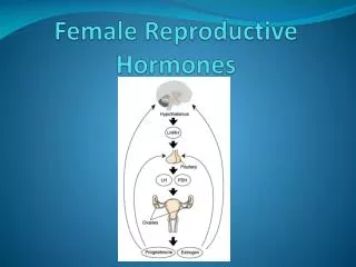
Female Anatomy and Hormones • Proestrus • Timing: approx 3 days before heat • Prostaglandin is released by the uterus into the bloodstream • Ovary contains a corpus luteum (CL) that produces progesterone • Prostaglandin causes the CL to regress lowering the levels of progesterone in the blood stream
Female Anatomy and Hormones • Proestrus • Declining Progesterone cause the pituitary gland to secrete FSH and LH • Higher FSH causes the ovaries to develop a follicle –fluid filled sac around the egg • The follicle releases estrogen in response to the FSH • Estrogen is what is responsible for behavioral changes in the female prior to heat.
Female Anatomy and Hormones • Proestrus • LH rates increase the day prior to estrus
Female Anatomy and Hormones • Estrus • Short period of time: 8-30 hours – most tend to cycle short • Signs of Estrus: • Standing to be bred • Mounting • Bellowing • Restlessness • Excitability • Mucus discharge • Reddish and swollen vuvla
Female Anatomy and Hormones • Estrus • Follicle releases the egg 10-14 hours after onset of estrus • Ovulation - Marks day 1 of the estrous cycle • Infundibulum catches the egg and transports it to the oviduct • If animal has been bred, sperm should be present in the fallopian tubes to fertilize the eggs (fertilization) • Follicle collapses after ovulation- leaving a mark on the ovary
Female Anatomy and Hormones • Metestrus • Follows ovulation: lasts 3-4 days • Estrogen declines and LH causes the former follicle to create a Corpus Luteum (CL) – • CL produces progesterone • Stimulates the uterus to prepare to nourish the embryo • The embryo continues to travel down the fallopian tubes towards the uterus
Female Anatomy and Hormones • Diestrus (if pregnant) • Embryo moves into the uterus and implants (conception) • CL produces large amounts of progesterone to stimulate the growth of the lining of the uterus • If pregnant, the uterus will not release prostaglandin and the CL will not regress • Progesterone is essential to maintaining the pregnancy
Female Anatomy and Hormones • Diestrus – (If open) • Lasts 12-15 days • Without pregnancy the uterus will release prostaglandin and the animal will renter proestrus
Show Me What You Know… • With a partner…create a “cycle” chart that shows what occurs during estrous • Must be included: • 4 Stages of Estrous • Hormones • Where these hormones are created Be creative when making this chart! 50 Points will be given for this assignment – Most creative will earn 25 extra bonus points!