Download
1 / 113
1.63k likes | 3.15k Vues
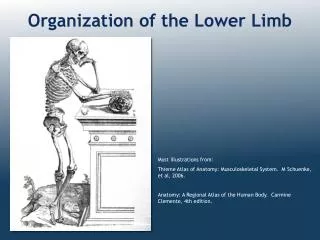
THE ANATOMY OF THE LOWER LIMB. BY DR. AHMAD KAMIL SHAHWAN PH.D. GENERAL SURGERY. THE LOWER LIMB ANATOMY. THE BONES OF THE LOWER LIMB : THE HIP BONE : It is a large bone ,thick in some places & thin in others.

E N D
THE ANATOMY OF THE LOWER LIMB BY DR. AHMAD KAMIL SHAHWAN PH.D. GENERAL SURGERY
THE LOWER LIMB ANATOMY THE BONES OF THE LOWER LIMB : THE HIP BONE : It is a large bone ,thick in some places & thin in others. It is formed of 3 bones : THE ILIUM , THEISCHIUM & THE PUPIS which are fused together before birth. They also fused at the acetabulum. There is a large opening below the acetabulum called the obturatorforamen
THE HIP BONE : THE ILIUM: is the upper expanded part of the hip bone ;it is the largest part & consists of body & a large flat wing called the ala of the ilium . It has 3 borders ;The upper border called the iliaccrest & ant. & post. borders. The iliac crest lies between the ant .sup. Iliac spine & the post. Sup. Iliac spine. Its ant. 2/3 is thick & convex outward & has inner & outer lips with an intermediate rough area in between , while the post. 1/3 is thin & convex inward & has 2 sloping surfaces separated by bony ridge.A bony prominence called the tubercle of theiliac crest is on the outer lip 5 cm . behind the ant. Sup. Iliac spine.
THE HIP BONE : The ant. border begins at the ASIS below which there is a notch then ant. Inf. Iliac spine. The post. Border begins in the PSIS & below it there is post inf. Iliac spine then form the greater sciatic notch then be continuous with the post. border of the ischium. The ilium has 2 surfaces :the outer(=gluteal) surface & the inner (=pelvic) surface.
THE HIP BONE : THE ISCHIUM : forms the lower & the post. part of hip bone &consists of body , tuberosity & one ramus. the body :the ant .border of the body is continuous below with the upper border of the ramus & both form part of the wall of the obturator foramen. The post. border of the body is continuous above with the post border of the ilium forming the lower part of the greater sciatic notch ,then project to form the ischial spine. , & then form the lesser sciatic notch before it form the ischial tuberosity .
THE HIP BONE : The ischial ramus is continuous in front with the inf. Ramus of the pubis. The ischial tuberosity is a very strong piece of bone which project from the inf. pole of the body of the ischium . It divided to 4 parts : 1- part give origin to semimembransus M. 2-part give origin to semitendenosus & long head of biceps Mm. 3-part give origin to adductor magnus M. 4-part which we sit on& does not give origin to any M.
THE HIP BONE : THE PUBIS :forms the lower & ant. Part of the hip bone& has a body & 2 rami. The body : is flat triangular part which articulate with its fellow at the symphysis pubis. The body has 3 borders : The upper border called the pubic crest & it ends laterlaly by a projection called the pubic tubercle. The lateral border is very sharp & form boundary of obturator foramen.
THE HIP BONE : The sup. Ramus is triangular in shape while the inf. Ramus starts at the symphysis pubis & run obliquely downwards & laterally to join the ischial ramus & form together the conjoint (ischio-pubic ) ramus. THE ARTICULATION OF THE HIP BONE: 1- above & behind with the sacrum to form the sacro-iliac joint. 2- below & in front with the other hip bone at symphysis pubis. 3- through the acetabulum with the head of the femur to form the hip joint.
THE FEMUR It is the longest & strongest bone in the body It is formed of upper end , shaft (body) & lower end. THE UPPER END :consists of the head , the neck ,the greater trochanter & the lesser trochanter . THE HEAD: is less than 2/3 of sphere & faces upwards forwards & medially. In life it is covered by a cartilage except with central depression called the fovea where the legamentum teres is attached.
THE FEMUR THE NECK : is 5 cm long & connect the head with the shaft .It forms an angle (110-120) with the axis of the shaft . This angle is smaller ( i.e. more acute ) in the female (who has wide pelvis ) than in male .This angle is normally 160 in children. THE GREATER TROCHANTER: is a large quadrangular piece of bone lies at the lateral & upper part of the junction between the neck & the shaft. In its medial surface there is deep depression called the trochantricfossa .
THE FEMUR THE LESSER TROCHANTER: is a small pyramidal projection. The intertrochanteric line : connects the greater & lesser trochanters in front & continues below the lesser troch. as the spiralline on the upper part of the shaft . The intertrochanteric crest : is a rough ridge joins the 2 troch. behind. In the middle of the crest there is a bony prominence called the quadratetubercle.
Blood Supply Of The Head of The Femur 1- Blood ascending upwards from the shaft along the cancellous bone. 2- Blood from the Vv. In the capsule of the hip joint. 3- Blood from the artery in the ligamentum teres.
THE SHAFT OF THE FEMUR It is cylindrical in shape & be flattened posteriorly & downward. It is very slightly curved( convex) anteriorly . Along the middle of the shaft posteriorly there is rough ridge called linea aspera with 2 lips (lat. & med.). The lateral lip of linea aspera superiorly join the gluteal tuberosity which extends upward to the base of greater troch. The medial lip of linea aspera passes above to form the spiral line & ends in the intertroch. Line.
THE SHAFT OF THE FEMUR The pactineal line arises from the lesser troch. & runs down parallel to the medial lip of linea aspera. In the lower 1/3 of the shaft the lat. & med. Lips diverge from each other & continue down as the lateral & medial supracondylarlines to the back of the lat. & med. Condyles respectively. leaving between them a flat triangular area called poplitealsurface .The med. Supracondylar line ends below in the adductor tubercle.
THE LOWER END OF THE FEMUR It consists of 2 condyles (med. & lat.) & 2 epicondyles( med. & lat.) . The condyles are large bony masses (the lat. Is larger) .Posteriorly the 2 cond. are separated from each other by a wide deep intercondylar fossa while anteriorly the 2 cond. fused to form the articular(patellar) surface. The most prominent parts of cond. is the epicondyles where between them posteriorly is the popliteal surface.
The joints of the femur 1- The head articulate with the acetabulum to form the hip joint. 2- The 2 femoral condyles articulate with the 2 tibial condyles in the knee joint . 3- The ant. surface of the lower end articulates with the upper 2/3 of the post. surface of the patella.
THE PATELLA • The patella(=the knee cap) : It is a flat & the largest sesamoid bone in the body located in the tendon of quadriceps femoris M. in front of the lower end of the femur . • It is triangular in shape with a base (upper border ) & an apex (rounded lower tip )& 2 borders (medial & lateral) & 2 surfaces (ant. & post.).
THE PATELLA • The lower 1/3 of the post. surface is rough while its upper 2/3 is smooth & is called the articularsurface as it articulates with the patellar surface of the femur . • The vastus medialis & vastus lateralis Mm are attached to the medial & lateral borders of the patella respectively while the vastus intermedius & the rectus femoris are attached to its upper border ( the base ). • The patellar ligament is attached to the apex. These attachments made it stable &rarely dislocated.
THE BONES OF THE LEG They are THE TIBIA & THE FIBULA THETIBIA: It is the large , weight bearing ,medial bone of the leg .It articulates with the condyles of the femur & the head of the fibula above & with the talus & distal part of the fibula below. It consist of expanded upper end , shaft & smaller lower end . The upper end of the tibia :There are lateral &medial condyles of the tibia which articulate with the lat. & med. Condyles of the femur . The lat. & med. Menesci intervening . Between the 2 condyles is the intercondylareminance.
THE TIBIA On the lateral aspect of the lateral condyle ;there is circular articular facet for the head of the fibula .
THE TIBIA The shaft of the tibia is triangular in cross section with 3 borders &3 surfaces ;Its ant. & med. borders with medial surface between them are subcutaneous ; the ant. border is prominent & form the shinof the leg . At the junction of the ant. border & the upper end is the tibialtuberosity which receives the attachment of ligamentum patellae . The med. border becomes rounded below where it becomes continuous with the medialmalleolus.
THE TIBIA The lateral(=interosseous) border gives attachment to interosseous membrane . The upper post. Surface shows an oblique line called the soleal line for the attachment of soleus M .Below it there is vertical line which extend to the interosseous border . The lower end of the tibia is slightly expanded & on the inf. surface shows saddle shape articular surface for articulation with talus, the lower end is prolonged down ward & medially to form the medial malleolus.
THE TIBIA The lateral surface of medial malleolus articulate also with the talus , On the lat. Surface of the lower end there is a wide rough depression for articulation with the fibula . THE JOINTS OF THE TIBIA: 1- the upper end : 1-1-the upper surface of the tibial condyles with the femoral condyles in the knee joint. 1-2-the fibular facet articulate with the flat circular facet on the fibula in the sup. tibio-fibular joint. (synovial joint.) .
THE TIBIA 2- the lower end : 2-1- the inf. surface of its lower end & the smooth lateral surface of med. malleolus articulate with the talus in the ankle joint. 2-2- the fibular notch articulate with the lower end of the fibula in the inf. tibio- fibularjoint.(a fibrous jt.),
THE FIBULA It is a very thin, long bone on the lateral side of the tibia .It takes no part in the articualion in the knee joint ; but below it forms the lat. Malleolus of the ankle joint. It takes no part in transmission of body weight, but it provides attachments for Mm. It consists of upper end (head ), neck , shaft & lower end. The upper end of the fibula: 1- The head :It is the bulky upper end has aposterolateral projection called the styloid process in which the biceps tendon is inserted .
THE FIBULA The upper part of its medial surface has a circular flat articular process to articulate with the fibular facet on the lateral condyle of the tibia . 2- The neck :the constricted upper part . The shaft :Its long twisted bone covered all with Mm. except triangular area above the lat. malleolus which is subcutaneous .It has 3 surfaces & 3 borders . The lower end of the fibula :is called the lateral malleolus , Its flattened from side to side & more pointed & longer than the medial malleolus. Its lateral surface is subcutaneous & its medial smooth surface for articulation with the lateral surface of the talus .while its posterior surface has a shallow groove for the tendons of peroneus longus & brevis .
THE FIBULA The fibula has 3 functions : 1- gives origin to Mm. of the leg . 2- form part of the ankle joint . 3- the lower end form a pulley for the tendons of peroneal mm. The Mm. attached to the fibula: One M.,biceps, inserted in the head of the fibula. Three mm. arise from the medial surface : 1- Extensor digitorum longus :from the upper ¾. 2- Peroneus tertius :from the lower ¼ . 3- extensor hallucis longus :from the middle ½.
THE FIBULA Two Mm. arise from the lateral (peroneal) surface: 1-peroneus longus :from the upper 2/3 . 2-Peroneus brevis : from the lower 2/3 . Three Mm. arise from the post. (flexor )surface : 1- Soleus :from the back of the head & upper 1/3 2-Tibialis post. M. from the medial side of post. surface . 3- Flexor hallucis longus: from the lat. side of post. Surface.